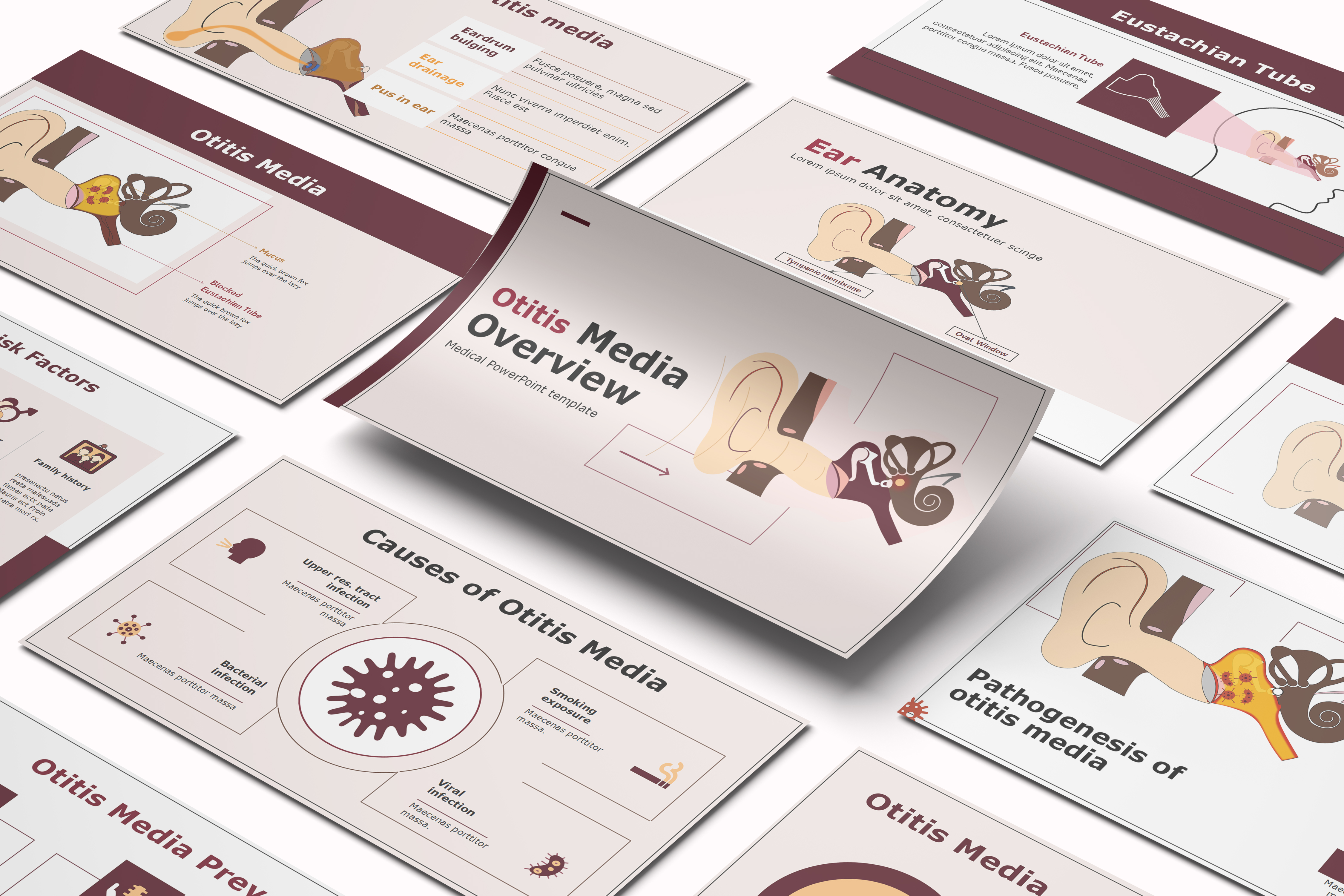
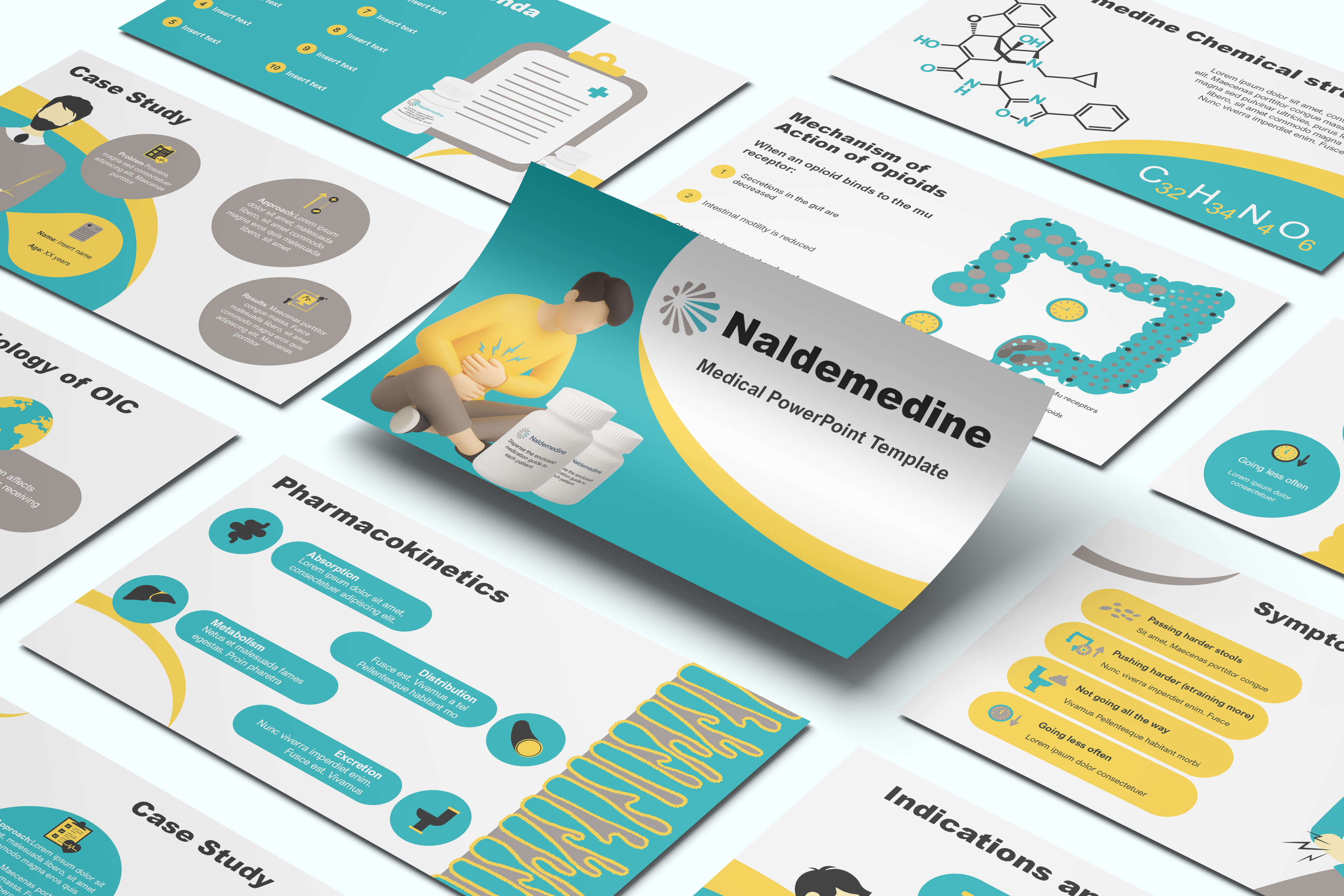
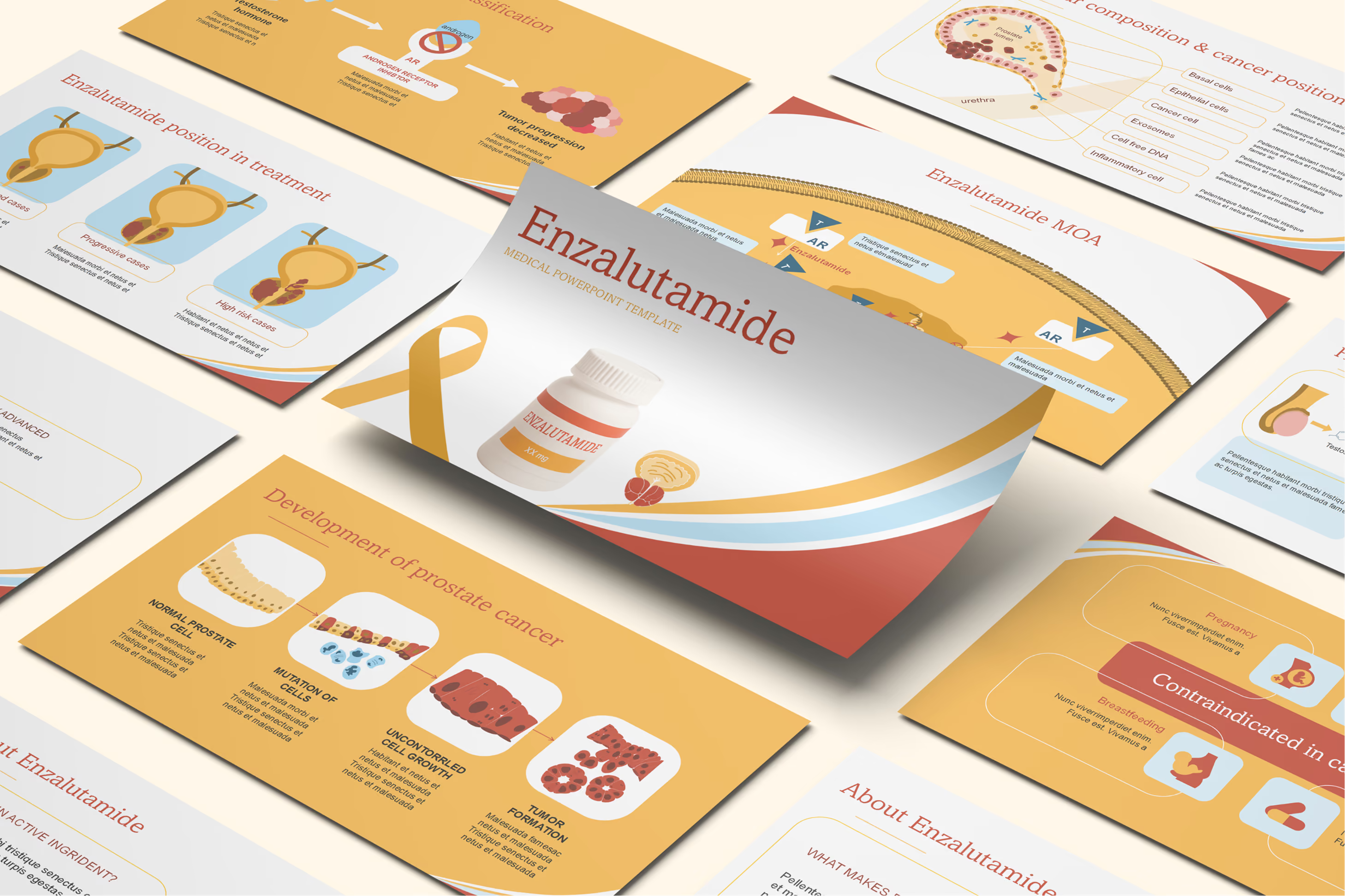
Glaucoma PowerPoint Template




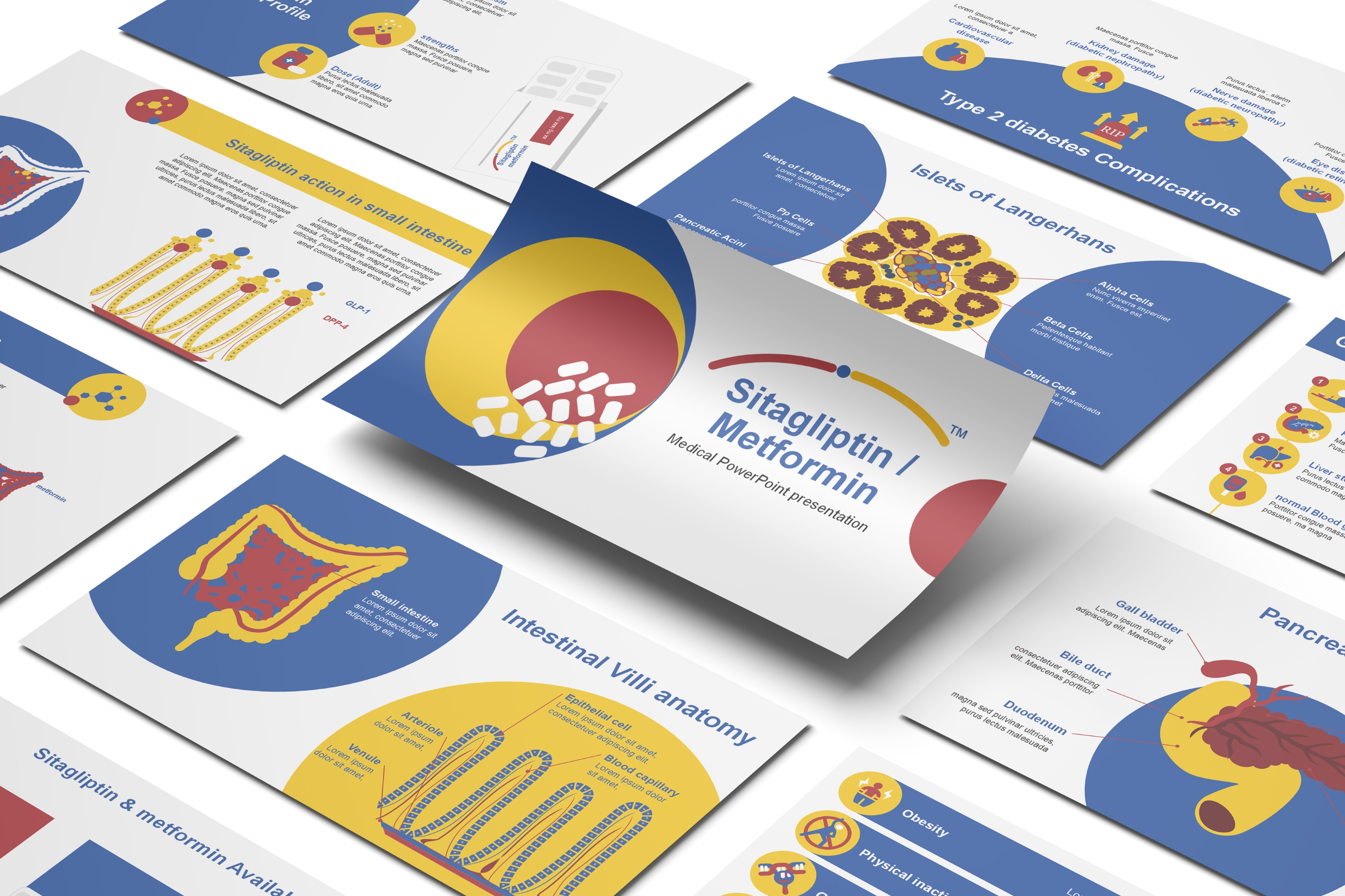
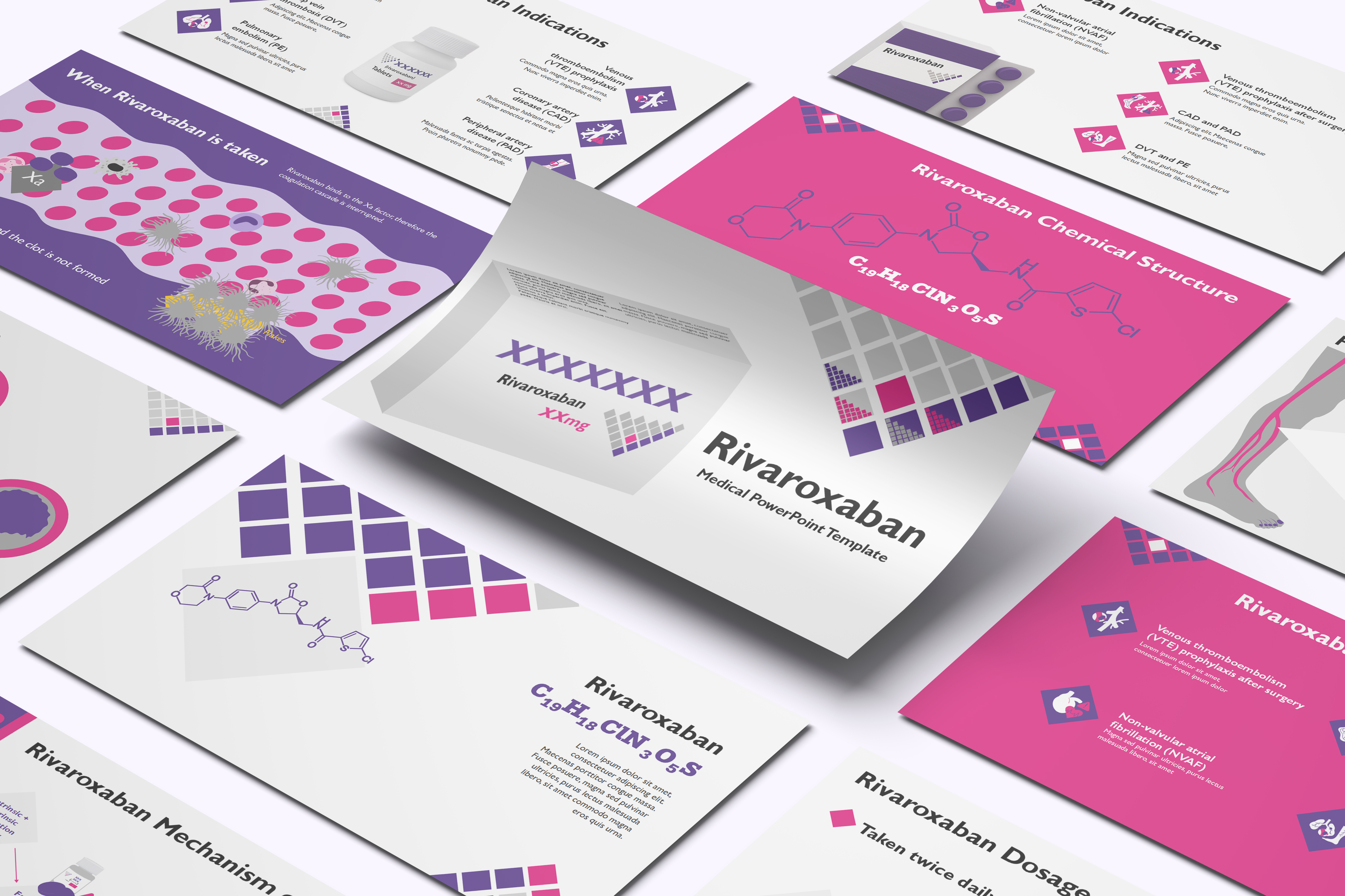
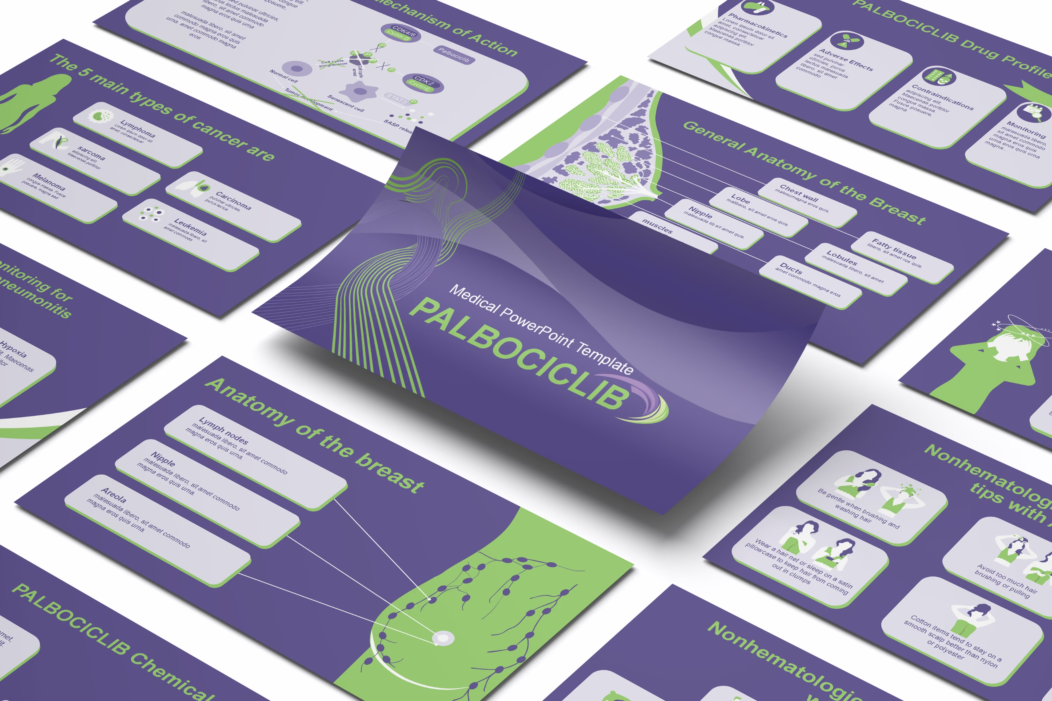
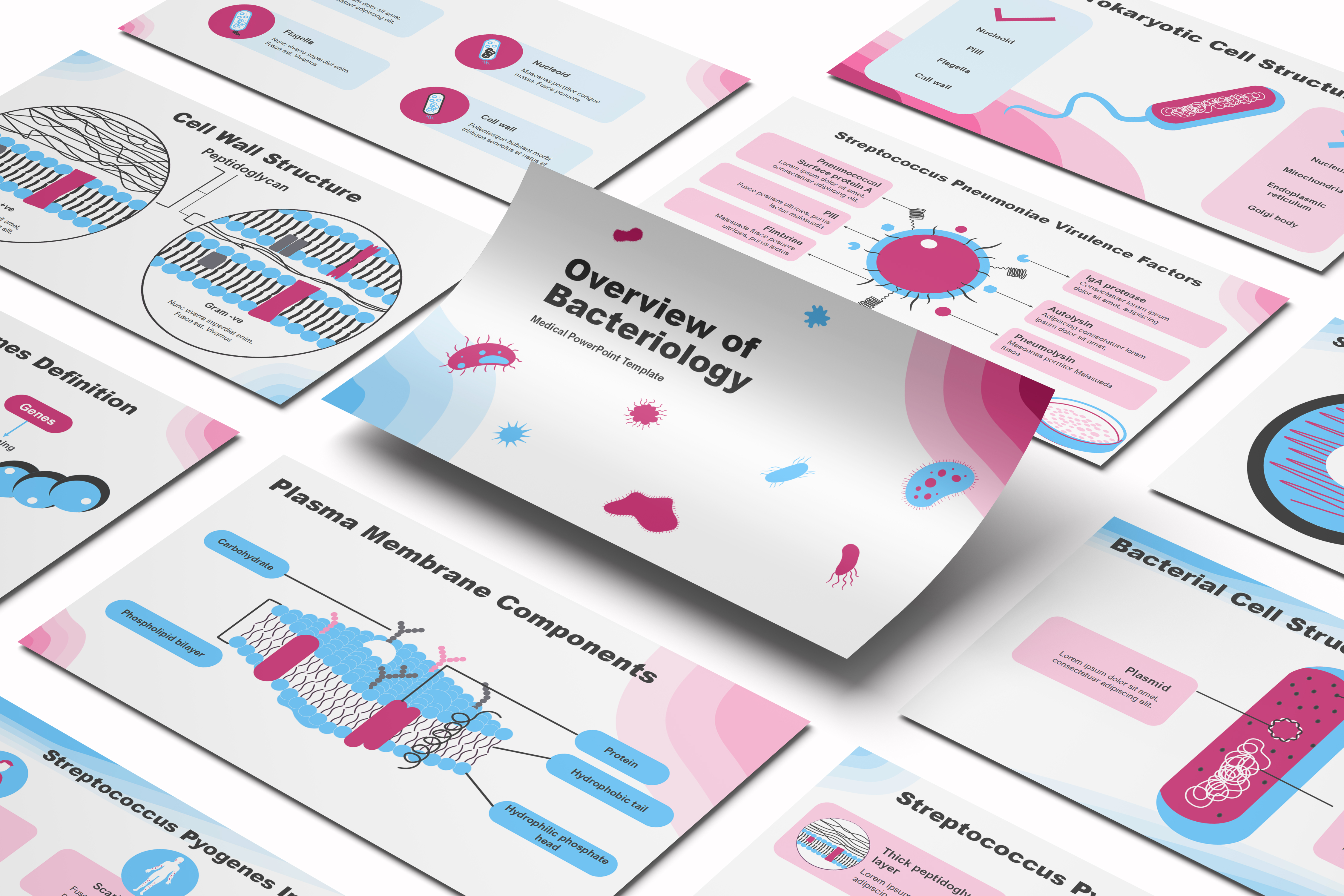
Template Preview
Designed for healthcare professionals creating clear and structured medical presentations.
Ideal for doctors, medical educators, and pharmaceutical teams preparing lectures, clinical education, product presentations, and training.
Fully editable in PowerPoint, allowing you to customize diagrams, charts, and slide layouts with your own medical content
Ready-to-use presentation structure that saves time and helps you build professional healthcare presentations quickly.
Fully Animated
Built-in animations help you present medical information clearly and make complex concepts easier to understand.
Editable Colors and Shapes
Editable colors and shapes help you easily customize the slides to match your content and presentation style, just drag and drop your content.
Professional medical layouts
Professional medical layouts help you organize clinical information, medical data, and treatment insights in a clear and structured way.

Created by Medical Professionals
This template prioritizes medical accuracy, clarity, and real-world usability. Each slide is designed with a medical audience in mind, helping you present clinical information, data, and treatment insights in a way that is structured, credible, and easy for healthcare professionals to understand.
Medical Training for Pharmaceutical & Life Sciences Companies
Ophthalmology training teams onboarding new sales reps can structure introductory sessions around this 50-plus-slide Glaucoma template's eye-anatomy foundation, optic nerve, ciliary body, and the three eye chambers, before moving into the aqueous humor drainage mechanism. New hires get a visual reference for how blocked or narrowed drainage produces open-angle versus angle-closure glaucoma, alongside the four-stage Mills staging system. The two-laser-procedure content, trabeculoplasty and peripheral iridotomy, gives trainees a structured reference for how surgical intervention differs by glaucoma type. New reps finish training able to connect the risk-factor profile, eye injury, age, family history, and diabetes, to why screening matters even before symptoms appear.
Medical Congress & Scientific Conference Presentations
Ophthalmologists preparing a conference session can build around this Glaucoma template's aqueous humor animation, tracing normal ciliary-body production and trabecular-meshwork drainage through the blockage mechanisms that raise intraocular pressure, rather than assembling ocular-physiology diagrams independently. The open-angle-versus-angle-closure comparison gives a speaker a ready framework for a mechanism-based glaucoma discussion. A Key Opinion Leader presenting at an ophthalmology conference can pair the existing four-stage Mills classification with their own institutional tonometry data. The two-laser-procedure content keeps the discussion clinically current for a specialist audience.
Pharmaceutical Product & Medical Affairs Presentations
Medical Affairs teams supporting a glaucoma medication or broader ophthalmology portfolio can use this Glaucoma presentation as disease-state groundwork for scientific exchange, covering aqueous humor physiology and the drainage-blockage mechanism before any product-specific data enters the conversation. Medical Science Liaisons can reference the open-angle-versus-angle-closure distinction when a scientific exchange conversation calls for context on why treatment approaches diverge by glaucoma type. The material stays at the disease-mechanism level rather than promoting a specific drug, so it still needs the customer's own medical and compliance review before Medical Affairs puts it in front of any audience. Ophthalmology and optometry teams gain a consistent scientific reference spanning mechanism, staging, and the full treatment continuum.
eLearning & SCORM Training Modules
Ophthalmology residency programs and optometry schools building a glaucoma module can customize this template with their own case studies, then convert the finished presentation into a SCORM-compliant module for their learning management system when self-paced study fits the curriculum better. The aqueous-humor and drainage-mechanism animations translate naturally into interactive checkpoints within a broader ophthalmology course. When a program needs a narrower scope, such as the staging system alone or a version tailored for optometry versus ophthalmology learners, the RxSlides team can adjust the template's coverage and branding. Beyond the current catalog, the RxSlides team can still design a presentation entirely from scratch to match a specific training goal.
Medical & Healthcare Education
Medical and optometry faculty teaching ophthalmology can use this template's eye-anatomy slides to establish the three-chamber structure before introducing how aqueous humor drainage failure raises intraocular pressure. The open-angle-versus-angle-closure framework gives students a clear structure for distinguishing the two major glaucoma mechanisms rather than treating the disease as uniform. The four-stage Mills classification connects classroom pathophysiology to how disease severity is actually documented in practice. Students leave with a structured understanding of glaucoma spanning anatomy, mechanism, staging, and treatment.
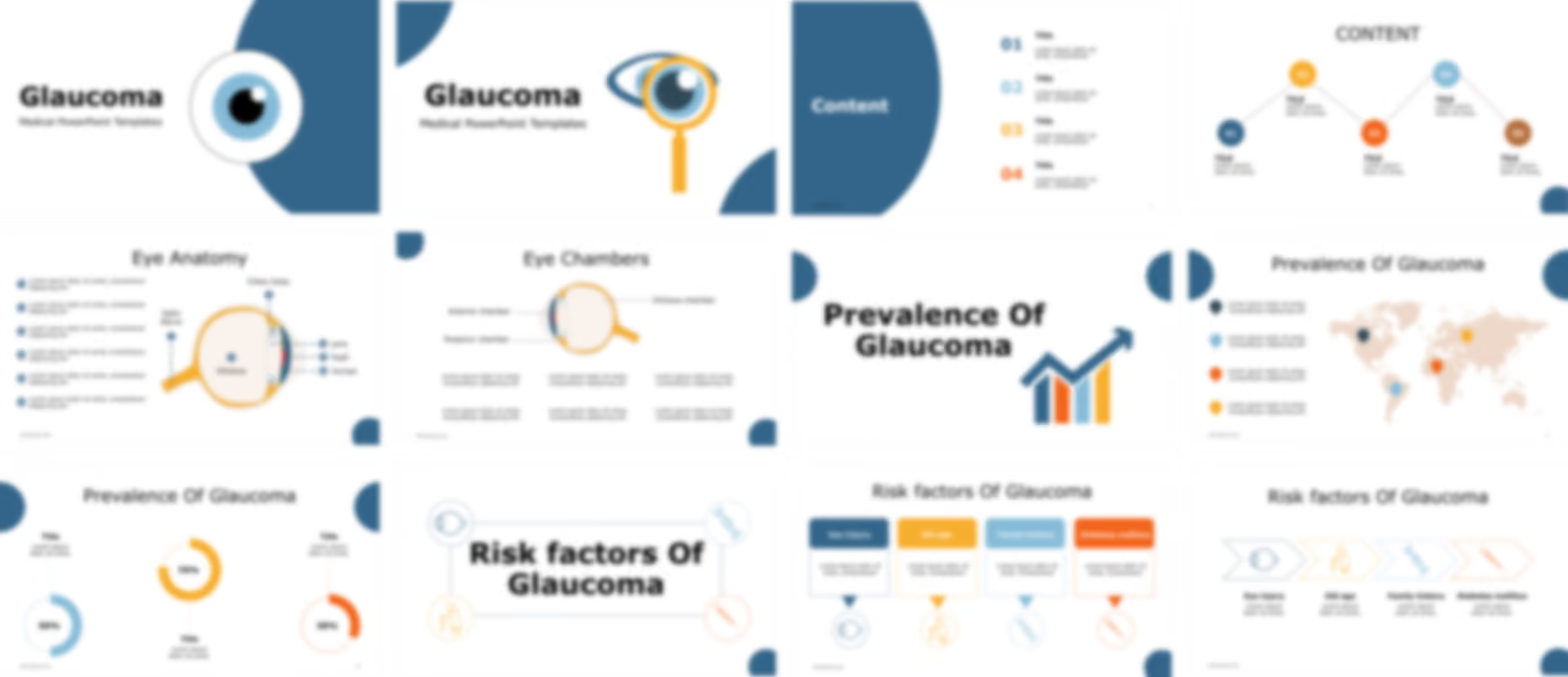
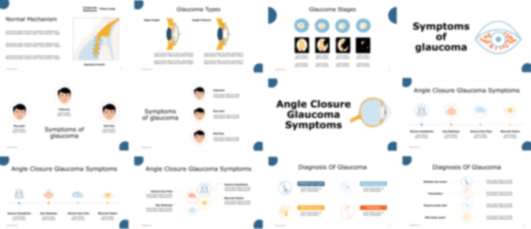
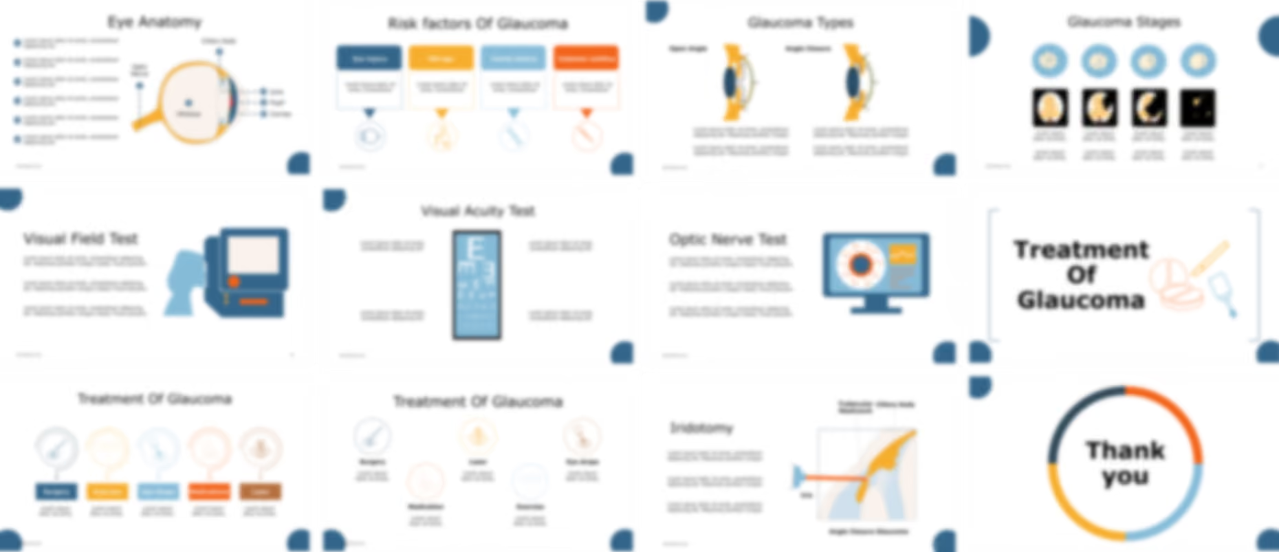
The Glaucoma PowerPoint template opens with cover slides and an editable table of contents before an anatomical illustration of the eye labels the optic nerve, vitreous, ciliary body, lens, pupil, cornea, and the anterior, posterior, and vitreous chambers. An epidemiology infographic then charts glaucoma's prevalence at both global and local levels, with percentages that can be updated to a specific dataset.
Medical icons cover risk factors including eye injury, old age, family history, and diabetes, before a definition slide describes glaucoma as optic nerve damage caused by fluid buildup that raises pressure inside the eye. Establishing the normal flow of aqueous humor through the ciliary body epithelium and trabecular meshwork first gives educators a shared reference point before an animated mechanism shows how blockage at the trabecular meshwork or narrowing of the drainage angle raises ocular pressure.
Comparative illustrations distinguish open angle and closed angle glaucoma and stage the disease from high pressure with no visual loss through early, moderate, and advanced glaucoma using the Mills grading system. Medical icons then visualize symptoms including severe headache, eye redness, severe eye pain, and blurred vision, in this editable medical presentation template.
Animated illustrations present diagnostic methods including dilated eye exams, visual acuity tests, slit lamp exams, and tonometry, before treatment content covers trabeculoplasty and laser peripheral iridotomy, explaining how each procedure creates an opening for aqueous humor to drain. The template closes with a set of customizable icons and illustrations for ophthalmology education.
Ready To Transform Your Presentation ?
Pay once, use forever