How to Present Bone Health: A Comprehensive Guide Using the Osteoporosis PowerPoint Template
Introduction: Pathophysiology and Asymptomatic Progression
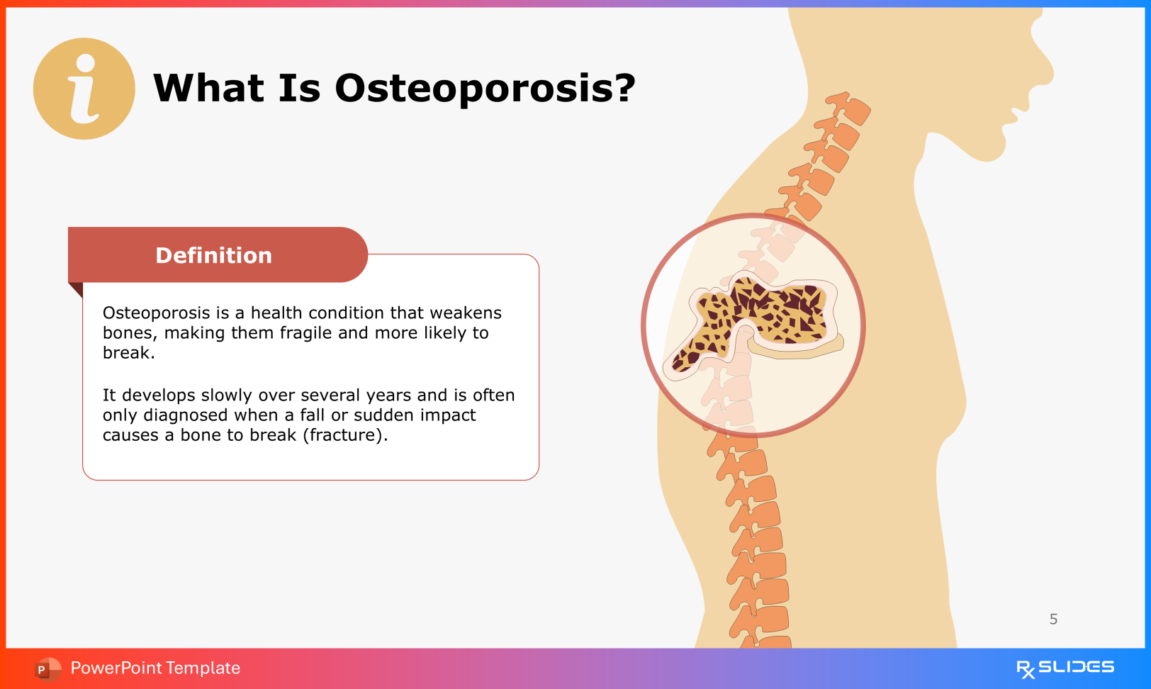
In clinical practice, osteoporosis is characterized by a systemic impairment of bone mass and micro-architecture that results in an increase in bone fragility and susceptibility to fractures. Unlike acute conditions with immediate symptomatic presentation, osteoporosis involves a progressive decline in Bone Mineral Density (BMD) that often goes undetected until a pathological fracture occurs.
For medical presenters using an Osteoporosis PowerPoint Template, the objective is to visualize this sub-clinical progression. The focus must be on the underlying metabolic imbalance—specifically, the rate of bone resorption exceeding bone formation—to emphasize the urgency of early screening and intervention.
Takeaway 1: Diagnostic Protocols
Addressing the Asymptomatic Phase
The primary clinical challenge of osteoporosis is its asymptomatic nature during the early stages of bone loss. Patients rarely present with pain or functional limitation until structural failure occurs.
Presentations should emphasize that the absence of symptoms does not correlate with skeletal health. The narrative must center on the Dual-Energy X-ray Absorptiometry (DEXA) scan as the gold standard for diagnosis. It is critical to define the diagnostic criteria: a T-score of -2.5 or lower confirms the presence of osteoporosis, necessitating therapeutic intervention regardless of the patient's fracture history.
Takeaway 2: Epidemiology and Disease Burden
Prevalence and Fracture Risk
To contextualize the clinical relevance of the disease, presenters should utilize current epidemiological data. The statistics highlight the significant morbidity associated with osteoporotic fractures:
Fracture Incidence: Approximately 50% of women and 25% of men will sustain an osteoporosis-related fracture in their lifetime.
Global Prevalence: The condition affects roughly one-third of postmenopausal women and one-fifth of men over the age of 50 worldwide.
Public Health Impact: With millions of adults currently diagnosed, osteoporosis represents a major burden on healthcare systems, primarily due to the costs and disability associated with hip and vertebral fractures.
Takeaway 3: Histopathology of Bone Loss
Cortical vs. Trabecular Micro-architecture
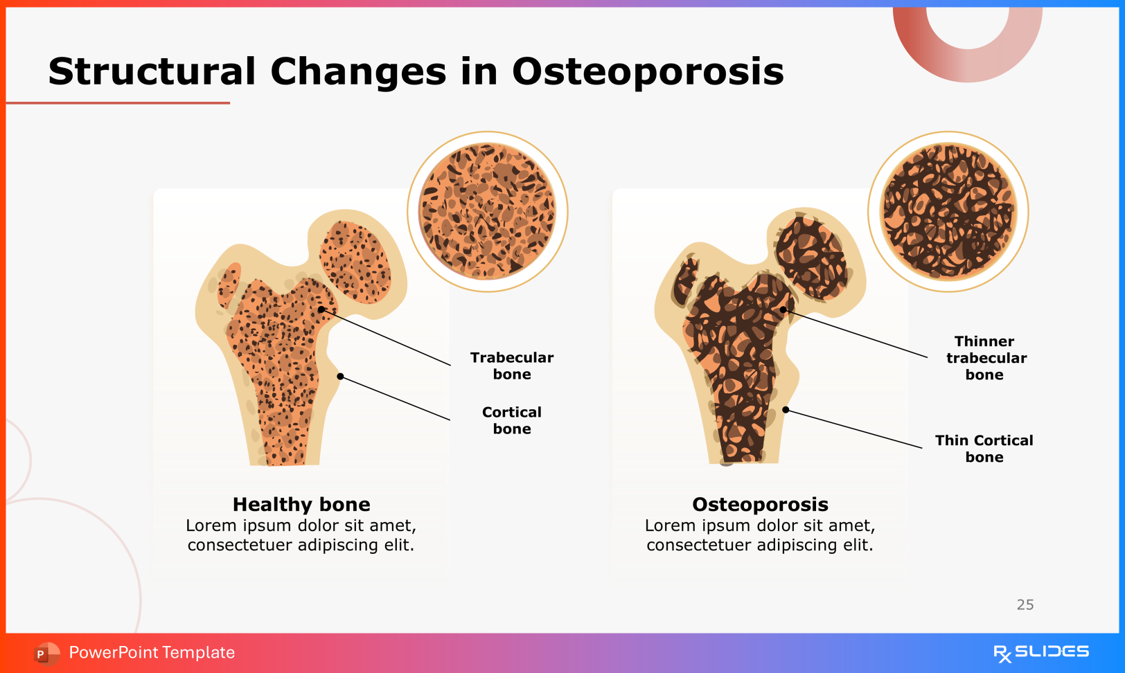
Osteoporosis is defined by specific histopathological changes. To accurately convey the severity of the disease, presentations must distinguish between the two primary types of bone tissue affected:
Trabecular (Cancellous) Bone: The internal network of trabeculae becomes thinned and disconnected, leading to a loss of structural connectivity.
Cortical (Compact) Bone: The outer protective layer undergoes thinning and increased porosity.
Visualizing the deterioration of the trabecular micro-architecture provides a clear physiological explanation for the reduction in mechanical strength and the increased risk of low-trauma fractures.
Takeaway 4: Endocrine Regulation of Remodeling
Estrogen Deficiency and the RANKL/OPG Pathway
A thorough medical presentation must address the cellular and hormonal mechanisms regulating bone homeostasis. The bone remodeling cycle is strictly controlled by the interaction between osteoblasts (formation) and osteoclasts (resorption).
Estrogen's Role: Estrogen promotes osteoblast survival and suppresses osteoclast activity.
The RANKL/OPG Ratio: Osteoblasts secrete Osteoprotegerin (OPG), which acts as a decoy receptor for RANKL. By binding to RANKL, OPG prevents it from activating the RANK receptor on osteoclasts, thereby inhibiting bone resorption.
In postmenopausal states, estrogen deficiency leads to a decrease in OPG relative to RANKL. This imbalance results in increased osteoclast differentiation and activity, accelerating bone resorption and net bone loss.
Takeaway 5: Prophylaxis and Risk Management
Modifiable and Non-Modifiable Risk Factors
Effective management requires a multifactorial approach to risk reduction. Presentations should outline the key pillars of prophylaxis:
Calcium and Vitamin D: Adequate calcium intake and Vitamin D status are essential for mineralization. Vitamin D promotes intestinal calcium absorption; deficiency leads to secondary hyperparathyroidism and increased bone turnover.
Lifestyle Factors: Smoking, excessive alcohol consumption, and physical inactivity are established risk factors that negatively impact BMD.
Weight-Bearing Exercise: Mechanical loading stimulates osteogenic activity, helping to maintain bone density.
Visual Showcase: Essential Slides for Clinical Presentations
To ensure the presentation is comprehensive and clinically rigorous, include the following structured slides:
Definition: Systemic skeletal disease characterized by low bone mass.
Etiology: Oxidative stress, chronic inflammation, and genetic predisposition.
Risk Stratification: Age, sex, glucocorticoid use, and lifestyle factors.
Epidemiology: Prevalence rates and fracture incidence statistics.
Anatomy: Periosteum, endosteum, and bone marrow structure.
Cellular Biology: Osteoblast and osteoclast function and interaction.
Histological Comparison: Normal vs. osteoporotic bone matrix.
Diagnostic Imaging: DEXA scan interpretation and T-score classification.
Why This Template is the Clinical Standard
This template is designed to meet the rigorous standards of medical education. A key feature is the inclusion of detailed renal physiology, specifically the mechanism of thiazide diuretics in the distal convoluted tubule (DCT).
By illustrating how thiazides inhibit the Na+/Cl− cotransporter (NCC) to enhance calcium reabsorption, the template supports advanced discussions on pharmacotherapy and calcium homeostasis. This level of detail establishes professional credibility and supports evidence-based teaching.
Conclusion
The management of osteoporosis relies on understanding the interplay between metabolic bone processes and structural integrity. By presenting the precise cellular mechanisms—from the RANKL/OPG pathway to trabecular thinning—clinicians can better educate patients on the necessity of early diagnosis and adherence to therapeutic regimens.
To deliver a presentation that aligns with these clinical standards, utilizing the RxSlides Osteoporosis PowerPoint Template ensures accuracy and professional impact.