A Guide to Presenting Wound Management in PowerPoint
Structuring High-Impact Clinical Presentations with Medical Visuals
The skin is the body’s largest organ and a critical immunological and mechanical barrier. When this barrier is disrupted by thermal injury, pressure-related ischemia, or surgical intervention, the resulting wound initiates a tightly regulated cascade of cellular and molecular events.
For healthcare professionals and medical educators, teaching wound management requires more than descriptive slides. It requires visual systems that connect clinical appearance, pathophysiology, and therapeutic decision-making across levels of care.
The Wound Management PowerPoint Presentation Template is designed to support this need, allowing educators to move seamlessly between anatomical context, biological mechanisms, and clinical intervention. Below is a structured guide on how to deploy this template effectively for professional medical education.
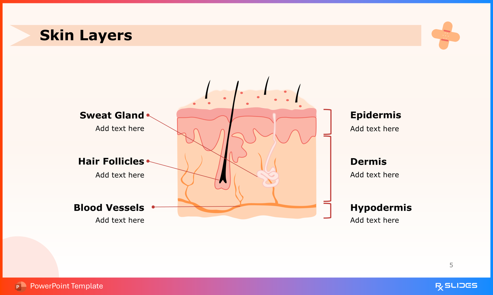
1. Skin Anatomy as the Foundation of Wound Classification
Effective wound education begins with precise anatomical orientation. Accurate identification of skin layers underpins burn classification, pressure injury staging, and surgical planning.
Use the Skin Layers slide to clearly delineate the epidermis, dermis, and hypodermis. This establishes shared terminology essential for interpreting injury depth, vascular involvement, and regenerative capacity.
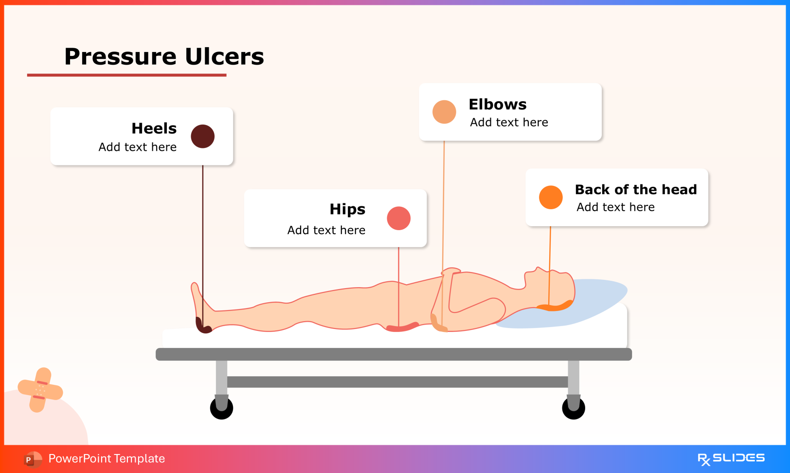
The Pressure Ulcers slides highlight high-risk pressure points, including the heels, hips, elbows, and occipital region, reinforcing the relationship between sustained pressure, ischemia, and tissue necrosis.
Use the Eschar slide to define eschar as firmly adherent necrotic tissue resulting from localized tissue death. The template supports discussion of etiologies such as ischemia, infection, gangrene, and toxin-mediated injury.
4. Debridement Strategies in Clinical Practice
Removal of non-viable tissue is central to effective wound management and infection control.
Mechanical debridement: Demonstrate the use of water to remove dead and other unwanted tissues
Biological debridement: Illustrates the controlled use of sterile larvae to selectively remove necrotic tissue
Autolytic debridement: Demonstrates the use of occlusive dressings to promote endogenous enzymatic breakdown
These slides support evidence-based decision-making based on wound type and patient condition.
5. Pharmacologic Agents and Advanced Dressings
Adjunctive therapies play a key role in optimizing wound healing outcomes.
Topical Pharmacologic Agents
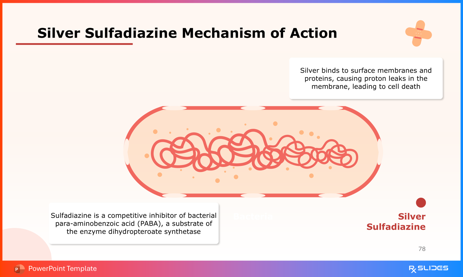
Silver sulfadiazine: Demonstrates silver-mediated disruption of bacterial cell membranes and sulfadiazine-induced inhibition of folic acid synthesis in organisms such as E. coli and Pseudomonas
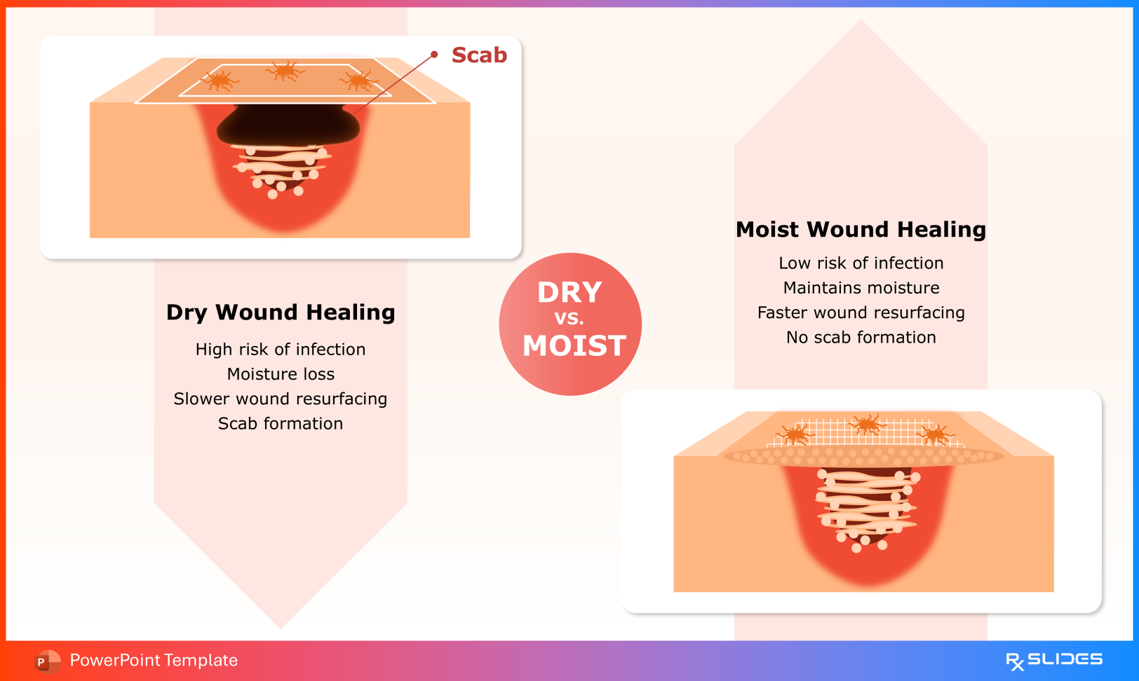
Use the dry vs. moist slide to compare dry, and moist gauze, emphasizing moisture balance and wound environment optimization.
Conclusion: From Clinical Observation to Cellular Insight
This template enables educators to bridge visible wound characteristics with underlying biological processes. It supports transitions from epidemiological considerations, such as pressure ulcer prevalence, to practical clinical interventions, including pharmacologic therapy and dressing selection.
For healthcare professionals, it offers a structured, visually coherent framework for teaching wound management across specialties and levels of care—without oversimplification.