Diabetic Retinopathy PowerPoint Template


No items found.
Diabetic Retinopathy PowerPoint Template: Animated Medical PowerPoint
- RxSlides presents a Diabetic Retinopathy PPT template to uncover the complexity of this topic in a straightforward, visually appealing way with the help of engaging animations.
- It could be difficult to understand the complex pathophysiology of diabetic retinopathy. Still, with this medical PowerPoint presentation, you can explain retinopathy pathology with Animation, ensure medical accuracy with the medical professionals at RxSlides and engage, educate and hold attention with visually compelling animations that transform passive learning into an active experience.
- Foster deeper understanding and knowledge retention in your audience.
- If you want to represent complex eye diseases, explore our Ophthalmology PowerPoint Templates collection with pre-animated eye illustrations for common eye conditions.
Diabetic Retinopathy PowerPoint Template Preview
Diabetic Retinopathy PowerPoint Template Content
Slide 1 - Title Slide (Diabetic Retinopathy)
.avif)
- Introduces the presentation on Diabetic Retinopathy.
- Presents a professional and visually descriptive design.
- Features a large, central illustration of a cross-section of a human eye with damaged vessels.
- Includes a blood glucose meter reading "126" for immediate context.
Slide 2 - Table of Contents (Navigation)
.avif)
- Provides a Table of Contents to outline the entire presentation structure.
- Utilizes six numbered eye icons as visual markers for each content area, enhancing navigation.
Slide 3 - Diabetic Retinopathy Introduction

- Serves as a smooth transition from the Table of Contents to the main content.
- Features a central icon combining a circle (representing the eye) and the glucose meter, visually linking the disease and its diabetic cause.
Slide 4 - ICD-10 and Key Details
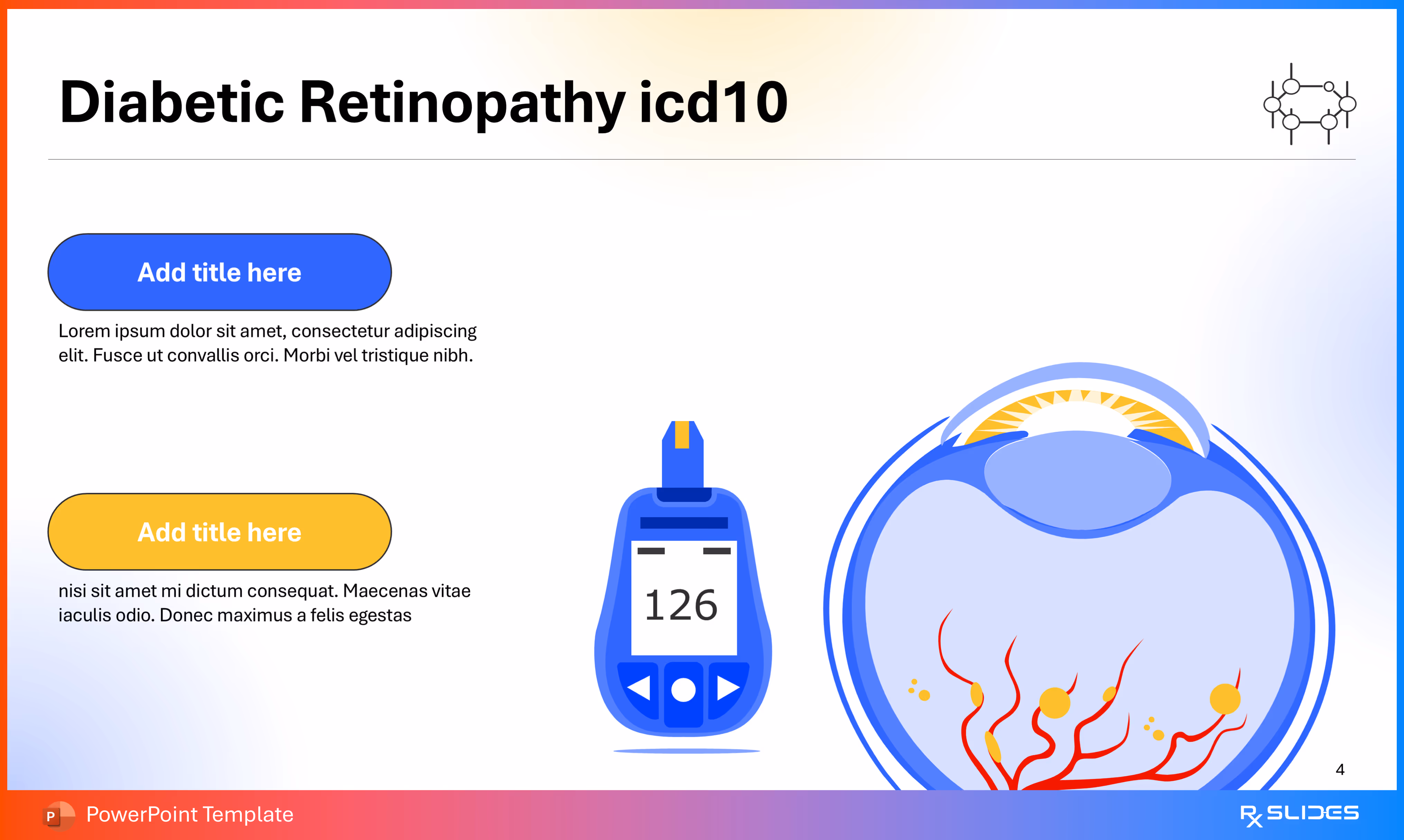
- Presents two distinct content points, such as the ICD-10 code, definitions, or symptoms.
- The layout uses two customizable text boxes (one blue and one yellow-orange) for easy comparison or separation of key data points.
Slide 5 - Vision Process Explanation
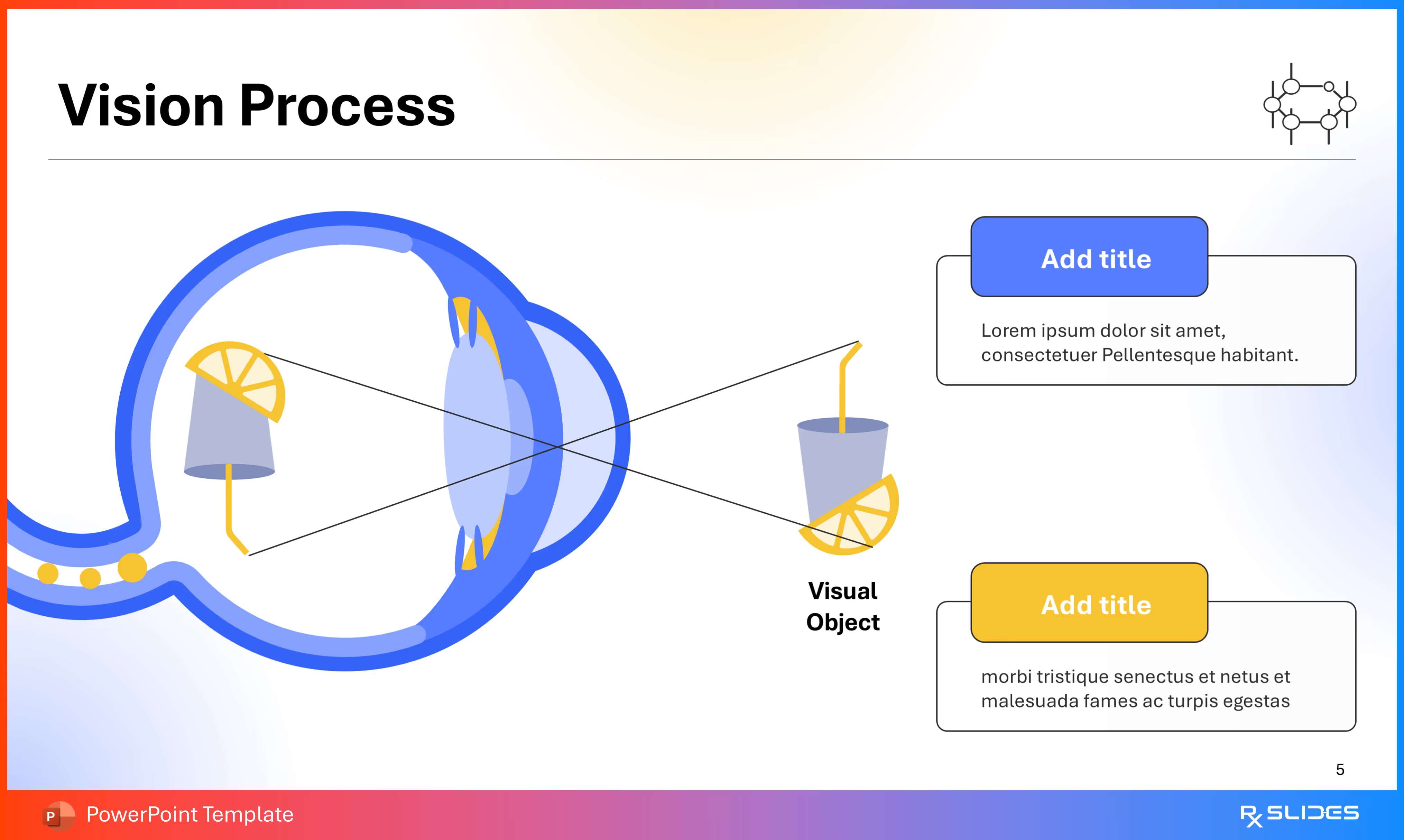
- Explains the Vision Process using an easy-to-understand illustration of the eye and light path.
- Features a fun analogy where light focuses through the lens and is captured at the back of the eye.
Slide 6 - Anatomy of The Eye (Labeling)
.avif)
- Describes five key parts of the eye's anatomy, such as the Iris, Lens, Pupil, Cornea, and Sclera.
- Features a large, simple diagram of the eye with lines pointing from labeled yellow boxes to the corresponding structures.
Slide 7 - Anatomy of The Eye (Internal Structures)
.avif)
- Describes five additional internal structures of the eye's anatomy, such as the Retina, Choroid, Ciliary Body, Anterior Chamber, and Vitreous Body.
- Features a large, simple diagram of the eye with lines pointing to the corresponding internal structures.
Slide 8 - Anatomy of The Eye (Posterior & Vascular)
.avif)
- Describes five crucial elements of the back of the eye, including the Macula, Optic Nerve, Arteries, and Veins.
- Features a large, simple eye diagram highlighting the posterior structures and the branching red and blue vessels.
Slide 9 - Diabetic Retinopathy Overview (Comparison)
.avif)
- Contrasts a Normal eye with one affected by Diabetic Retinopathy side-by-side.
- The design uses two eye diagrams, one showing healthy vessels and the other showing damaged vessels, separated by a "VS" icon.
Slide 10 - Diabetic Retinopathy Introduction (Retinal View)
.avif)
- Illustrates the difference between a Normal retina and one affected by Diabetic Retinopathy at a detailed level.
- The design uses two circular views, displaying a healthy retinal vessel network versus a network showing hemorrhages and damage.
Slide 11 - Normal Retina (Detailed View)
.avif)
- Shows the healthy anatomy of the Normal Retina, specifically highlighting the vascular and structural components.
- Features a stylized illustration of an eye exam, with a magnified, labeled view of the retina identifying structures like the Macula, Fovea, Artery, and Vein.
Slide 12 - Retina in Diabetic Retinopathy (Pathology)
.avif)
- Demonstrates and labels the pathological features found in a retina affected by Diabetic Retinopathy.
- Features a magnified, labeled view of the diseased retina, highlighting key signs like Hemorrhages, Hard Exudates, Aneurysm, Abnormal blood vessels, and Cotton Wool Spots.
Slide 13 - Diabetic Retinopathy Prevalence (Section Divider)
.avif)
- A Divider slide for a smooth transition to the critical section on the Prevalence and global impact of Diabetic Retinopathy.
- Features a central icon combining the eye motif with a globe.
Slide 14 - Prevalence of Diabetic Retinopathy (Statistics)
.avif)
- Showcases key statistics, highlighting the high rate of Diabetic Retinopathy among people with diabetes.
- Features a prominent 25% pie chart segment next to a large glucose meter.
Slide 15 - Progression of Diabetic Retinopathy (3 Stages)
.avif)
- Illustrates the three stages of progression for Diabetic Retinopathy, making complex disease development easy to understand.
- Features three numbered eye diagrams, showing a gradual increase in vessel damage and hemorrhages from stage 1 to stage 3.
Slide 16 - Diabetic Retinopathy Causes (Section Divider)
.avif)
- A Divider slide to transition to the crucial section detailing the Causes of Diabetic Retinopathy.
- Features a central icon combining the eye motif with stylized, damaged cells.
Slide 17 - Causes of Diabetic Retinopathy (Blood Sugar Focus)
.avif)
- Explains how uncontrolled blood sugar is the primary cause of damage in Diabetic Retinopathy.
- Features a large, detailed illustration of the damaged retina and a person testing their blood sugar with a glucose meter.
- Powerfully illustrates the link between high glucose levels and retinal damage.
Slide 18 - Causes of Diabetic Retinopathy (Glucose Detail)
.avif)
- Shows the specific relationship between excess Glucose and damage to the Retina, detailing the underlying mechanism.
- Features a large, simple cross-section of the eye showing vessel damage, paired with two separate text boxes for specific content points.
Slide 19 - Diabetic Retinopathy Risk Factors (Section Divider)
.avif)
- A Divider slide to transition to the important section detailing the Risk Factors for developing Diabetic Retinopathy.
- Features a central icon combining the eye motif with a silhouette of a person.
- Ensures your presentation structure is easy to follow and professionally introduces a new topic segment.
Slide 20 - Diabetic Retinopathy Risk Factors (Overview)
.avif)
- Presents six major risk factors that increase the likelihood of developing Diabetic Retinopathy.
- Features a central eye icon surrounded by six separate, relevant icons—such as a blood drop, a pregnant figure, and a cholesterol symbol—each linked to a key factor like High Blood Pressure or Family History.
Slide 21 - Risk Factors Detail (Grid Layout)
.avif)
- Provides detailed explanations for each of the six major risk factors.
- Presents the six risks (like High Blood Pressure, Race, High Cholesterol, Diabetes, Family history, and Pregnancy) in a clean, professional three-by-two grid.
Slide 22 - Risk Factors Presentation (Eye Diagram Focus)
.avif)
- Summarizes the six major risk factors using an abstract, artistic arrangement of eye-related diagrams.
- Features two central eyes connected to a third circular diagram filled with yellow dots.
Slide 23 - Diabetic Retinopathy Symptoms (Section Divider)
.avif)
- A Divider slide to transition to the crucial section detailing the Symptoms experienced by patients.
- Features a central icon combining the eye motif with a stylized, open eye graphic.
Slide 24 - Symptoms of Diabetic Retinopathy (Visual Icons)
.avif)
- Presents six key symptoms associated with Diabetic Retinopathy.
- Features a central eye icon and six surrounding blue circles, each with an icon representing a specific symptom, such as Blurred Vision, Eye Pain, or Floaters.
Slide 25 - Clinical Signs of Advanced Retinopathy (Detailed)
.avif)
- Describes the specific clinical signs seen in advanced Diabetic Retinopathy, making complex pathology easy to teach.
- Features a large, magnified view of the retina with four lines pointing to and labeling key lesions like Microaneurysms, Dot and blot hemorrhages, and Retinal edema.
Slide 26 - Clinical Signs of Advanced Retinopathy (Additional Signs)
.avif)
- An essential continuation slide to label and describe four additional, critical signs of advanced Diabetic Retinopathy.
- Highlights signs including Cotton wool spots, Macular edema, intraretinal microvascular abnormalities, and Neovascularization.
Slide 27 - Diabetic Retinopathy Pathophysiology (Section Divider)
.avif)
- A Divider slide to transmit the presentation to the complex section detailing the Pathophysiology.
- Features a central icon combining the eye motif with a detailed illustration of the damaged retinal vessels.
Slide 28 - Pathophysiology of Diabetic Retinopathy (Mechanisms)
.avif)
- Explains the underlying biological mechanisms by focusing on two key problems: Increased Vascular Permeability and Intra retinal Microvascular Abnormalities.
- Features a large, simple diagram of the eye showing damaged blood vessels, corresponding to the detailed descriptions in the red text boxes.
Slide 29 - Normal vs. Diabetic Capillaries (Cellular Detail)
.avif)
- Contrasts the cellular structure of a Normal capillary against a Damaged capillary found in Diabetic Retinopathy.
- Illustrates a healthy capillary with intact Pericytes versus a diseased one showing cellular damage and degenerating pericytes.
Slide 30 - Normal vs. Diabetic Capillaries (Advanced View)
.avif)
- Contrasts a Normal capillary against a Damaged capillary from a retina affected by Diabetic Retinopathy.
- Pairs a microscopic view of a healthy, stable capillary with a detailed pathological view showing immune cells, inflammation, and cellular breakdown.
Slide 31 - Pathophysiology Flowchart (Complete Process)
.avif)
- Illustrates the entire progression of Diabetic Retinopathy, starting from the root cause (Diabetes and Blood Sugar).
- Moves sequentially through four stages, linking the initial problem to Oxidative Stress and culminating in the final clinical stages: NPDR (Non-proliferative) and PDR (Proliferative).
Slide 32 - Pathophysiology Flowchart (Multiple Pathways)
.avif)
- A Flowchart that illustrates how Diabetes initiates multiple damaging pathways, including Hyperlipidemia, hormones, hyperglycemia, and Hypertension, all leading to Diabetic Retinopathy.
- Tracks four primary inputs from the glucose meter through Oxidative Stress, showing the development of retinal damage.
Slide 33 - Diabetic Retinopathy Key Signs (Summary)
.avif)
- A Summary slide to visually reinforce five of the most critical clinical signs of Diabetic Retinopathy in an easy-to-read grid.
- Features five separate, circular icons showing retinal damage like Hard Exudates, Hemorrhages, and Newly Formed Blood Vessels.
Slide 34 - Diabetic Retinopathy Key Signs (Visual Summary)
.avif)
- A Summary slide to visually reinforce five of the most important clinical signs of Diabetic Retinopathy (an alternative to slide 33).
Slide 35 - Diabetic Retinopathy Stages (Section Divider)
.avif)
- A Divider slide to transmit the presentation to the essential section detailing the different Stages of Diabetic Retinopathy progression.
- Features a central icon combining the eye motif with a detailed illustration of damaged retinal vessels.
Slide 36 - Stages of Diabetic Retinopathy (Progression)
.avif)
- Illustrates the three distinct stages of Diabetic Retinopathy progression.
- Features three numbered eye diagrams, showing the disease advance from No Apparent stage to Non-proliferative (early damage) and finally to Proliferative (advanced damage).
Slide 37 - The Retina in Stages of Diabetic Retinopathy (Cross-Section)
.avif)
- Visualizes the microscopic tissue changes that occur in the retina across the three stages of progression.
- Features three horizontal cross sections, illustrating the change from a healthy surface to the presence of exudates and damage, and finally to bleeding and new vessel growth.
Slide 38 - Non-Proliferative vs. Proliferative Retinopathy (Final Comparison)
.avif)
- Provides a visual and textual breakdown of the two advanced stages: Non-Proliferative and Proliferative Diabetic Retinopathy.
- Features two detailed eye cross-sections, labeling the specific lesions for each stage, such as Microaneurysm in NPDR and Retinal Neovascularization in PDR.
Slide 39 - Non-Proliferative Diabetic Retinopathy (Signs)
.avif)
- Presents three key early clinical signs of Non-Proliferative Diabetic Retinopathy (NPDR).
- Features a detailed eye cross-section illustrating early damage like Hemorrhage, Microaneurysm, and Cotton wool spots.
Slide 40 - Background Retinopathy (No Treatment Needed)
.avif)
- Details the characteristics of Background Retinopathy, emphasizing that it typically does not require immediate treatment.
- Features three distinct icons: a detailed retinal view, an eye with a checkmark indicating Sight is not affected, and a syringe with an 'X' indicating No treatment needed.
Slide 41 - Pre-Proliferative Retinopathy (Vision at Risk)
.avif)
- Details the characteristics of Pre-Proliferative Retinopathy, emphasizing that vision is now at risk.
- Features a retinal view showing Bleeding into the retina, an eye with a warning sign indicating Vision is at risk, and a clipboard signaling Frequent screening.
Slide 42 - Proliferative Diabetic Retinopathy (Signs)
.avif)
- Presents two key advanced clinical signs of Proliferative Diabetic Retinopathy (PDR).
- Features a magnified view illustrating advanced damage like Vitreous hemorrhage and Retinal neovascularization (new, abnormal blood vessel growth).
Slide 43 - Diabetic Retinopathy Diagnosis (Section Divider)
.avif)
- A Divider slide to transition to the essential section detailing the Diagnosis methods.
- Features a central icon combining the eye motif with a classic slit lamp exam device.
Slide 44 - Diagnosis of Diabetic Retinopathy (4 Key Tests)
.avif)
- Outlines the four key methods used to diagnose Diabetic Retinopathy.
- Lists four crucial diagnostic steps: Comprehensive dilated eye exam, Fundus photography, Optical coherence tomography (OCT), and Fluorescein angiography (FA).
Slide 45 - Diagnosis of Diabetic Retinopathy (Test Layout)
.avif)
- Outlines the four key methods used to diagnose Diabetic Retinopathy, using a clean, symmetrical layout.
- Lists four crucial diagnostic steps: Comprehensive dilated eye exam, Fundus photography, Optical coherence tomography (OCT), and Fluorescein angiography (FA).
Slide 46 - Comprehensive Dilated Eye Exam (Visual Explanation)
.avif)
- Explains the process and purpose of the Comprehensive Dilated Eye Exam.
- Illustrates an Un Dilated Pupil versus a Dilated Pupil, showing how dilation allows doctors to see the back of the eye more clearly.
Slide 47 - Fundus Photography (Visual Comparison)
.avif)
- Explains the process and diagnostic utility of Fundus Photography.
- Includes two magnified comparison circles showing a Normal retina (healthy vessels) and a retina with Diabetic Retinopathy (damaged vessels).
Slide 48 - Optical Coherence Tomography (OCT)
.avif)
- Explains the process of Optical Coherence Tomography (OCT), an essential, non-invasive imaging test.
- Features an illustration of a patient undergoing an OCT scan, with a magnified view of the retina.
Slide 49 - Fluorescein Angiography (FA) (Step-by-Step)
%20(Step-by-Step).avif)
- Explains the three-step process of Fluorescein Angiography (FA).
- Features a clear, sequential illustration showing: Step 1 (injection of the dye), Step 2 (dye travel), and Step 3 (the resulting detailed image).
Slide 50 - Diabetic Retinopathy Complications (Section Divider)
.avif)
- A Divider slide to smoothly transition to the critical section on Complications.
- Features a central icon combining the eye motif with a stylized bursting star/cross-hatch pattern.
Slide 51 - Diabetic Retinopathy Complications (Flowchart)
.avif)
- Illustrates the four most severe Complications that can result from advanced Diabetic Retinopathy.
- Features a sequential breakdown leading to four major outcomes: Diabetic macular edema, Neovascular glaucoma, Retinal detachment, and ultimately Blindness.
Slide 52 - Diabetic Retinopathy Complications (Visual Summary)
.avif)
- An alternative slide (51) that highlights four major outcomes: Retinal detachment, Blindness, Neovascular glaucoma, and Diabetic macular edema.
- All complications are linked to the central image of damaged retinal vessels.
Slide 53 - Retinal Detachment (Complication Detail)
.avif)
- Provides a text box for the detailed complication of Retinal Detachment.
- Features a large, cross-sectional illustration of the eye, highlighting where the retina has pulled away from the underlying tissue.
Slide 54 - Neovascular Glaucoma (Complication Detail)
.avif)
- Provides a text box for the detailed complication of Neovascular Glaucoma.
- Features an illustration of the eye, highlighting the link between damaged retinal vessels at the back of the eye and issues in the front of the eye.
Slide 55 - Diabetic Macular Edema (Complication Detail)
.avif)
- Provides a text box for the detailed complication of Diabetic Macular Edema (DME).
- Features an illustration of the eye, specifically highlighting the swollen macula area with yellow coloring and damaged vessels.
Slide 56 - Diabetic Retinopathy Treatment (Section Divider)
.avif)
- A Divider slide to transmit the presentation to the essential section detailing the Treatment options.
- Features a central icon combining the eye motif with a syringe positioned over the damaged retina.
Slide 57 - Diabetic Retinopathy Therapy (4 Main Options)
.avif)
- Outlines the four main treatment options for Diabetic Retinopathy.
- Organizes treatments into four categories: Laser treatment, Eye injections, Steroid eye implants, and Eye surgery.
Slide 58 - Diabetic Retinopathy Therapy (Checklist Summary)
.avif)
- Summarizes the four main treatment options using a clear, vertical checklist design.
- Lists treatments like Laser treatment, Eye injections, Eye surgery, and Steroid eye implants, each paired with an icon illustrating the procedure.
Slide 59 - Laser Treatment (Photocoagulation Comparison)
.avif)
- Compares the two main types of Laser Treatment: Photocoagulation and Pan-retinal photocoagulation (PRP).
- Features a side-by-side illustration showing a focused laser beam for localized damage versus widespread laser application (PRP).
Slide 60 - Laser Treatment (Surgical Detail)
.avif)
- Explains the process and goal of Laser Treatment using a direct visual of the procedure.
- Features an illustration of the eye showing a bright Laser Beam being aimed at the damaged area, with the treated area visible as a cluster of yellow spots (laser burns).
Slide 61 - Steroid Eye Implants (Procedure Detail)
.avif)
- Explains the procedure and purpose of Steroid Eye Implants.
- Features an illustration showing a syringe carefully implanting a slow releasing medication (like Dexamethasone) into the vitreous body.
Slide 62 - Eye Injections (Anti-VEGF Agents)
.avif)
- Explains the procedure and purpose of Eye Injections (focusing on Anti-VEGF agents).
- Features an illustration showing a syringe carefully injecting a yellow substance (representing the Anti-VEGF medicine) into the vitreous body.
Slide 63 - VEGF Receptors Mechanism of Action (Cellular Detail)
.avif)
- Explains the cellular mechanism of action for Anti-VEGF therapy.
- Features a diagram showing the VEGF protein (Vascular Endothelial Growth Factor) binding to its Receptors on a cell membrane.
Slide 64 - Anti-VEGF Agents Mechanism of Action (Drug Binding)
.avif)
- Explains how Anti-VEGF Agents work at the cellular level.
- Features a detailed diagram showing the drug (Anti-VEGF Agents) intercepting and binding to the VEGF protein before it can activate its Receptors.
Slide 65 - Vitrectomy Surgery (Procedure Detail)
.avif)
- Explains the procedure and purpose of Vitrectomy Surgery.
- Features an illustration showing surgical instruments (Cannula, Light, and Vitrectomy device) inserted into the eye to remove blood (Hemorrhage).
Slide 66 - Diabetic Retinopathy Prevention (Section Divider)
.avif)
- A Divider slide to smoothly transmit the presentation to the essential section detailing the Prevention strategies.
- Features a central icon of an eye with damaged vessels that are crossed out (symbolizing stopping the disease).
Slide 67 - Prevention of Diabetic Retinopathy (6 Key Actions)
.avif)
- Outlines the six most important Prevention strategies for Diabetic Retinopathy.
- Uses six circular icons representing blood sugar control, regular exercises, smoking cessation, hypertension control, lipid control, and weight management.
Slide 68 - Retinal Screening (Action Steps)
.avif)
- Outlines the final action steps related to Retinal Screening.
- Features a central illustration of the damaged retina surrounded by four numbered text boxes for key instructional points on screening frequency, preparation, and follow-up.
Slide 69 - Thank You (Final Slide)
.avif)
- Concludes the presentation with a thank you slide.
- Features a large Snellen Eye Chart with the words "Thank You" overlaid, next to a stylized eye showing the progression of Diabetic Retinopathy.
Features of the Template
- 100% editable PowerPoint template.
- Editable colors, you can change according to your presentation style and company branding guidelines.
Purchase Template
Purchase the fully editable PowerPoint open file template
Template Specifications
Slides count:
60+ Slides
Free monthly updates via email
Compatible with:
Microsoft PowerPoint
File type:
PPTX
Dimensions:
(16:9)