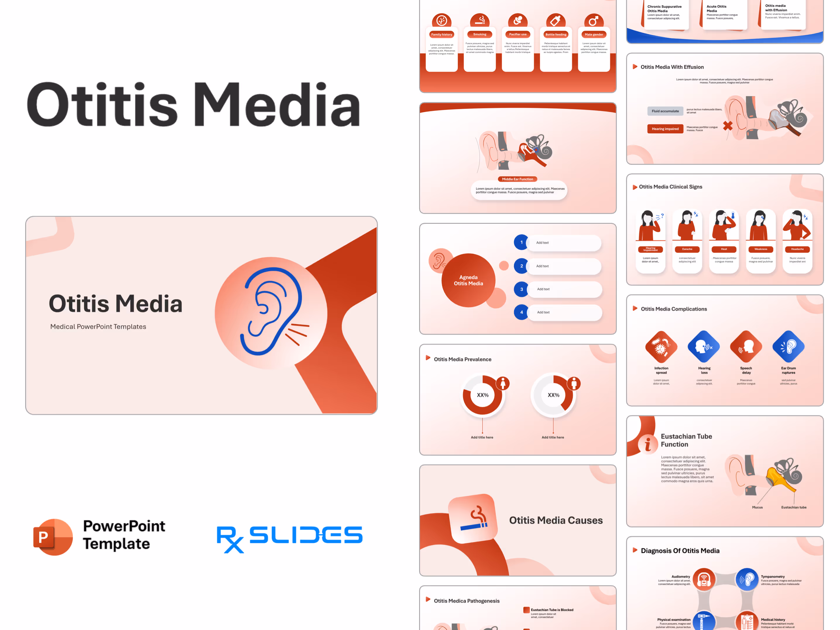
Otitis Media PowerPoint Template


No items found.
Otitis Media PowerPoint Template Preview
Otitis Media PowerPoint Template Content
Slide 1 - Otitis Media (Title Slide)
.avif)
- Title Slide for the presentation on Otitis Media.
- The design features a large, stylized illustration of the human ear in blue outline, with red sound/pain lines emanating from the inner part, symbolizing the condition.
Slide 2 - Agenda Otitis Media (Six-Point Overview)
.avif)
- Agenda slide or Table of Contents for the Otitis Media presentation.
- The design uses a central red circular icon of the ear to focus the agenda points around the topic.
- Six numbered points (1 through 6) are arranged around the central icon in a semi-circle flow, providing space for titles like "Definition," "Causes," "Symptoms," "Diagnosis," "Treatment," and "Prevention".
Slide 3 - Otitis Media Agenda (Four-Point Overview)
.avif)
- This slide is an alternative Agenda or Key Takeaways page.
- The design features a large central red circle displaying the title "Agneda Otitis Media" with a small ear icon to the left.
- Four numbered, rounded, white text boxes (1 through 4) are arranged vertically to the right.
Slide 4 - Otitis Media Anatomy (Section Title)
.avif)
- Section divider slide to transmit to Anatomy relevant to Otitis Media.
- The slide features a large, stylized blue ear icon on a pink, rounded-square shape, with red sound/pain lines.
- The title "Otitis Media Anatomy" is prominent on the right.
- The design uses a soft pink, red, and blue color scheme.
Slide 5 - Ear Anatomy (Structure Overview)
.avif)
- Explains the major parts of the ear relevant to Otitis Media.
- The slide features a large, detailed cross-section illustration of the ear, dividing it into three key sections: Outer ear, Middle ear (The location of Otitis Media), and Inner ear.
- Key anatomical features are highlighted, including the Tympanic membrane (eardrum) and the Oval Window.
Slide 6 - Middle Ear Function (Focus on Pathophysiology)
.avif)
- Details the structure and function of the Middle Ear.
- The slide features a large cross-section illustration of the Middle Ear on the top half. The illustration shows the tympanic membrane (eardrum) and the fluid-filled space containing the ossicles.
- A prominent callout box at the bottom is titled "Middle Ear Function".
Slide 7 - Eustachian Tube (Anatomy and Function)
.avif)
- Explains the Eustachian Tube, its location, and its critical role in draining the middle ear and equalizing pressure.
- The slide features a simplified illustration of the human head and ear anatomy, with a zoomed-in section focusing on the Eustachian Tube.
Slide 8 - Eustachian Tube in Adults and Children (Key Difference)
.avif)
- Illustrates the anatomical difference in the Eustachian Tube between Children and Adults.
- The slide features two simplified side-profile illustrations of the head.
Slide 9 - Eustachian Tube Function (Detailed View)
.avif)
- Provides a more detailed look at the function of the Eustachian Tube.
- The slide features a cross-section illustration highlighting the Eustachian Tube and its connection to the middle ear.
- The illustration specifically labels the Eustachian tube and Mucus.
- A key text box on the left, titled "Eustachian Tube Function,".
Slide 10 - Otitis Media Definition (Section Title)
.avif)
- Section divider slide to transmit to the Definition and potentially the Pathology of Otitis Media.
- The slide features a large, stylized blue ear icon on a pink, rounded-square shape, with a single red drop beneath the ear, symbolizing fluid or inflammation.
- The title "Otitis Media Definition" is prominent on the left.
Slide 11 - Otitis Media (Pathology Explained)
.avif)
- Illustrates the key pathological process in Otitis Media.
- The slide features a prominent illustration of the ear's cross-section on the right.
- The illustration clearly shows the middle ear space filled with a yellow fluid and labeled Mucus.
- The Blocked Eustachian Tube is explicitly labeled.
Slide 12 - Otitis Media Prevalence (Section Title)
.avif)
- Section divider slide to transmit to Prevalence and Epidemiology of Otitis Media.
- The slide features a large, stylized bar graph icon with a rising red arrow, visually representing statistics and increasing numbers.
- The title "Otitis Media Prevalence" is prominent on the right.
Slide 13 - Otitis Media Prevalence (Gender Comparison)
.avif)
- Compares the prevalence of Otitis Media between female and male populations.
- The slide features two prominent doughnut charts side-by-side.
- The left chart features a female icon (woman's head).
- The right chart features a male icon (man's head).
Slide 14 - Otitis Media Risk factors (Section Title)
.avif)
- Section divider slide to transmit to the Risk Factors for Otitis Media.
- The slide features a large, stylized blue and red caution/exclamation icon in a triangular shape.
- The title "Otitis Media Risk factors" is prominent on the left.
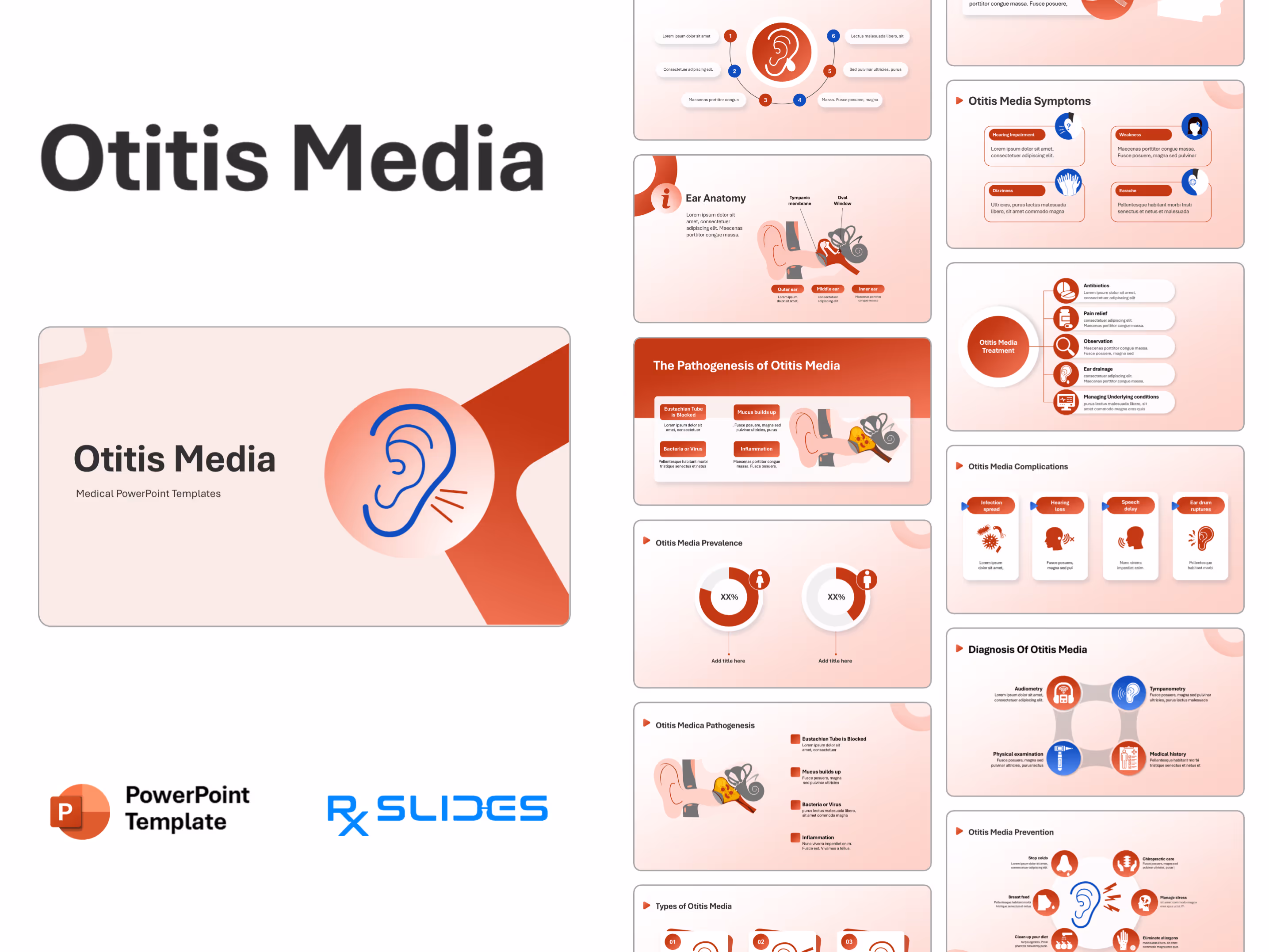
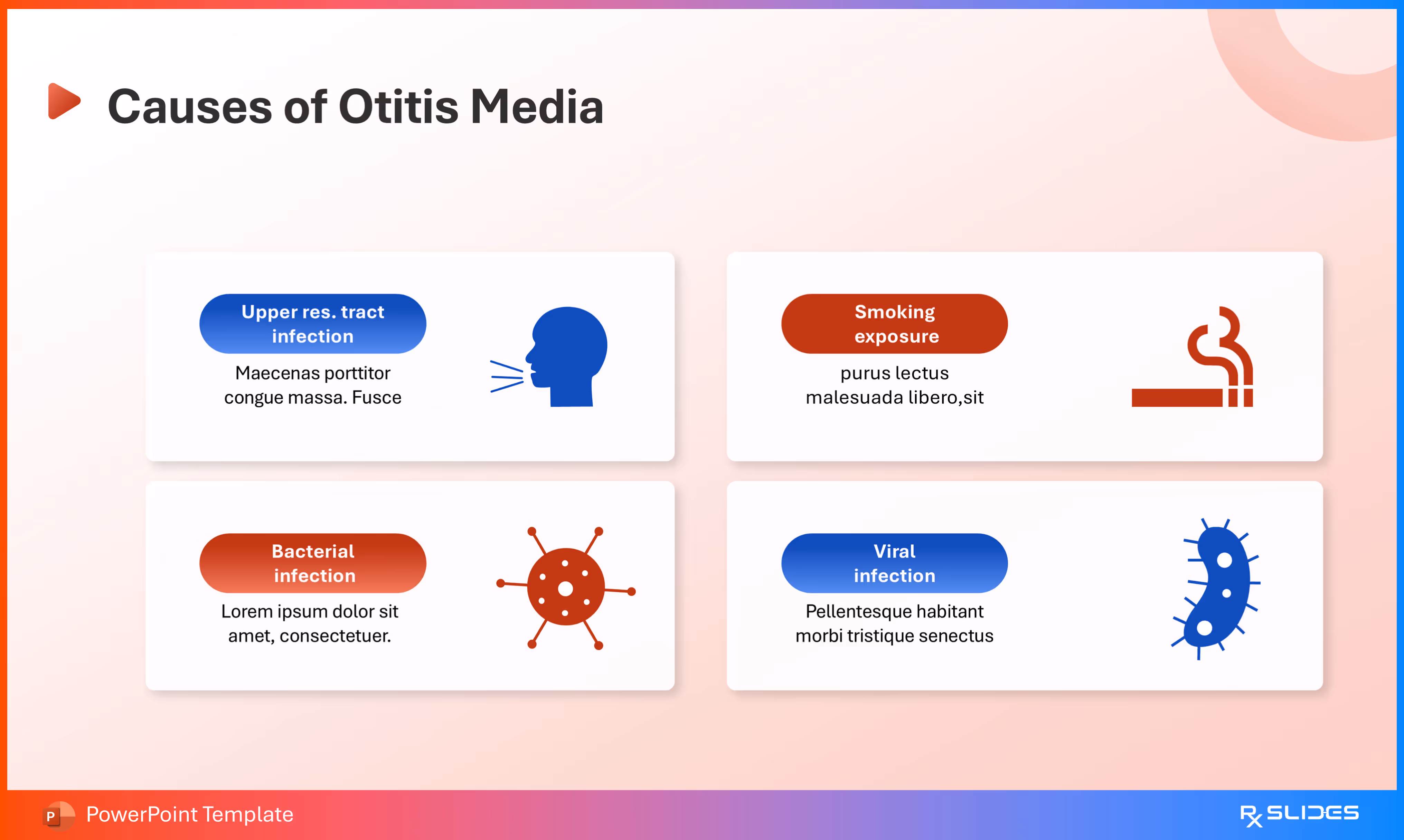
Slide 15 - Otitis Media Risk Factors (Five Key Factors)
.avif)
- Illustrates five key risk factors associated with developing Otitis Media.
- The design uses a flow chart of five alternating red and blue circular icons connected by curved lines, showing the progression or relationship between risk factors: Family history, Smoking, Pacifier use, Bottle feeding, and Male gender.
Slide 16 - Otitis Media Risk Factors
- Describes five key risk factors associated with developing Otitis Media.
- The design uses five vertical columns, each dedicated to a single risk factor and providing significant space for descriptive text beneath it.
- Each column includes a prominent icon and title: Family history, Smoking, Pacifier use, Bottle feeding, and Male gender.
Slide 17 - Otitis Media Causes (Section Title)
.avif)
- Section divider slide to transmit to Causes of Otitis Media.
- The slide features a large, stylized cigarette icon with smoke in a blue and red color scheme.
- The title "Otitis Media Causes" is prominent on the right.
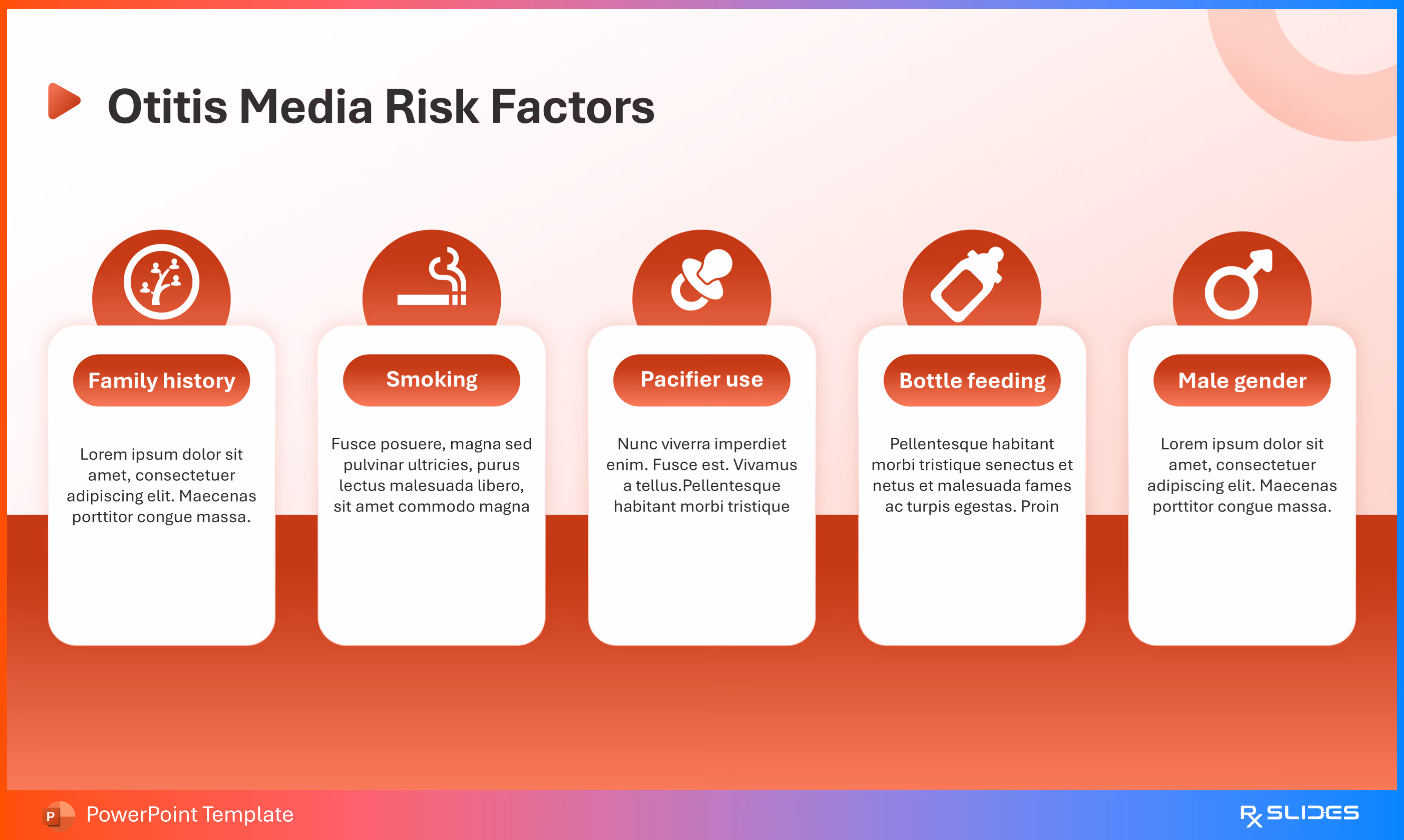
Slide 18 - Causes of Otitis Media
- Details the four main causes and contributing factors of Otitis Media.
- The design uses layout visually separating the causes into four distinct boxes, each with its own icon and color scheme (red and blue): Upper res. tract infection, Smoking exposure, Bacterial infection, and Viral infection.
Slide 19 - Otitis Media Pathogenesis (Section Title)
.avif)
- Section divider slide to transmit to Pathogenesis (mechanism of disease) of Otitis Media.
- The slide features a large, stylized icon showing multiple viral and bacterial pathogens in a blue and red color scheme.
- The title "Otitis Media Pathogenesis" is prominent on the left.
Slide 20 - Otitis Media Pathogenesis (Four Steps)
.avif)
- Explains the four steps in the development of Otitis Media.
- The slide features a central illustration showing the ear anatomy with the middle ear space filled with yellow fluid.
- Four key stages are listed vertically in red boxes on the right: Eustachian Tube is Blocked, Mucus builds up, Bacteria or Virus (Infection begins), and Inflammation.
Slide 21 - The Pathogenesis of Otitis Media
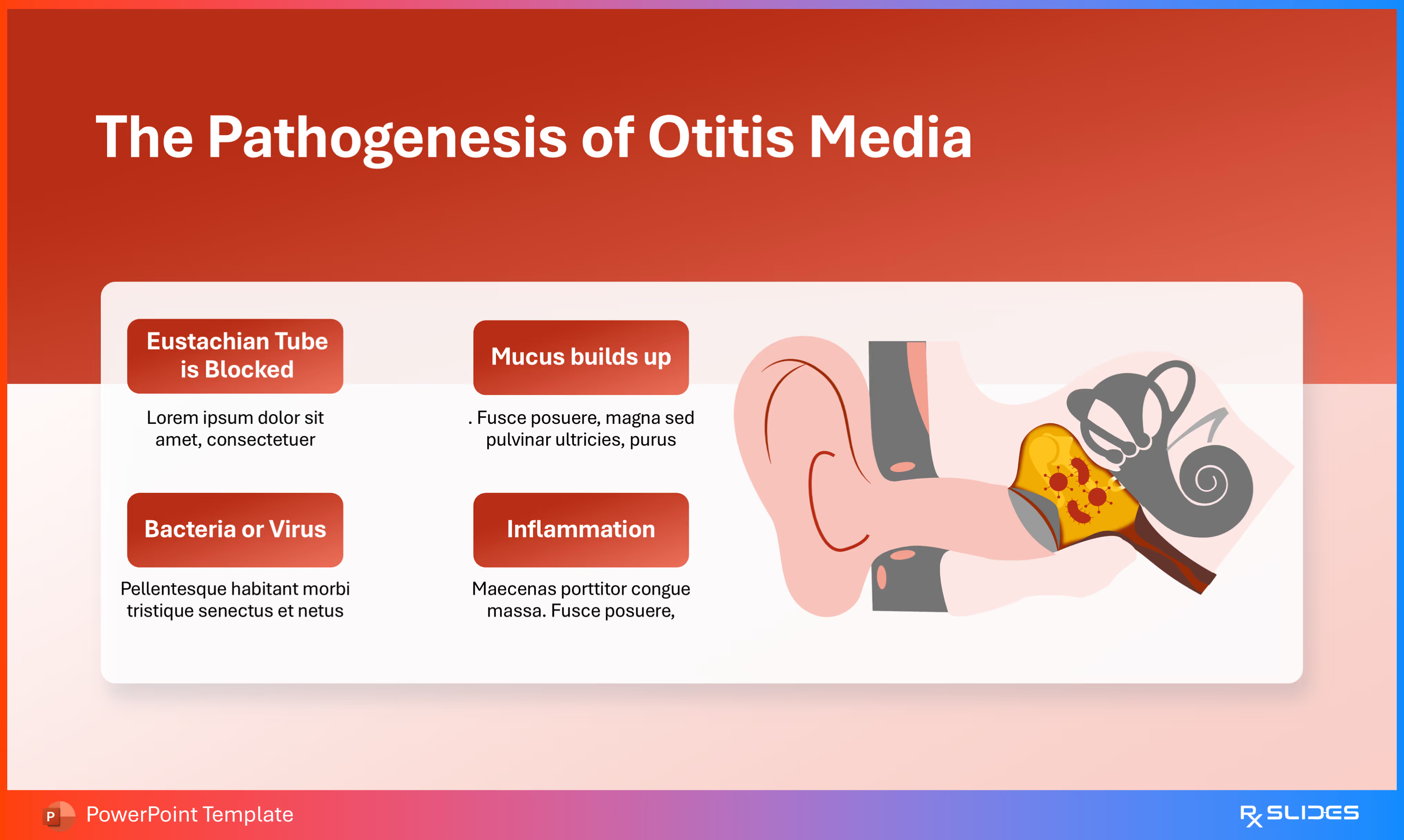
- Segments the four steps in the development of Otitis Media.
- The slide features a large illustration of the ear pathology on the right, showing the middle ear space filled with yellow, inflamed fluid and a blocked Eustachian tube.
- The four key stages are presented in a 2x2 grid of contrasting red boxes on the left, offering separate description space for each step: Eustachian Tube is Blocked, Mucus builds up, Bacteria or Virus, and Inflammation.
Slide 22 - Otitis Media Types (Section Title)
.avif)
- Section divider slide transmit to Types or classifications of Otitis Media.
- The slide features a large, stylized blue ear icon with red sound/pain lines.
- The title "Otitis Media Types" is prominent on the right.
- The design uses a soft pink, red, and blue color scheme.
Slide 23 - Types of Otitis Media
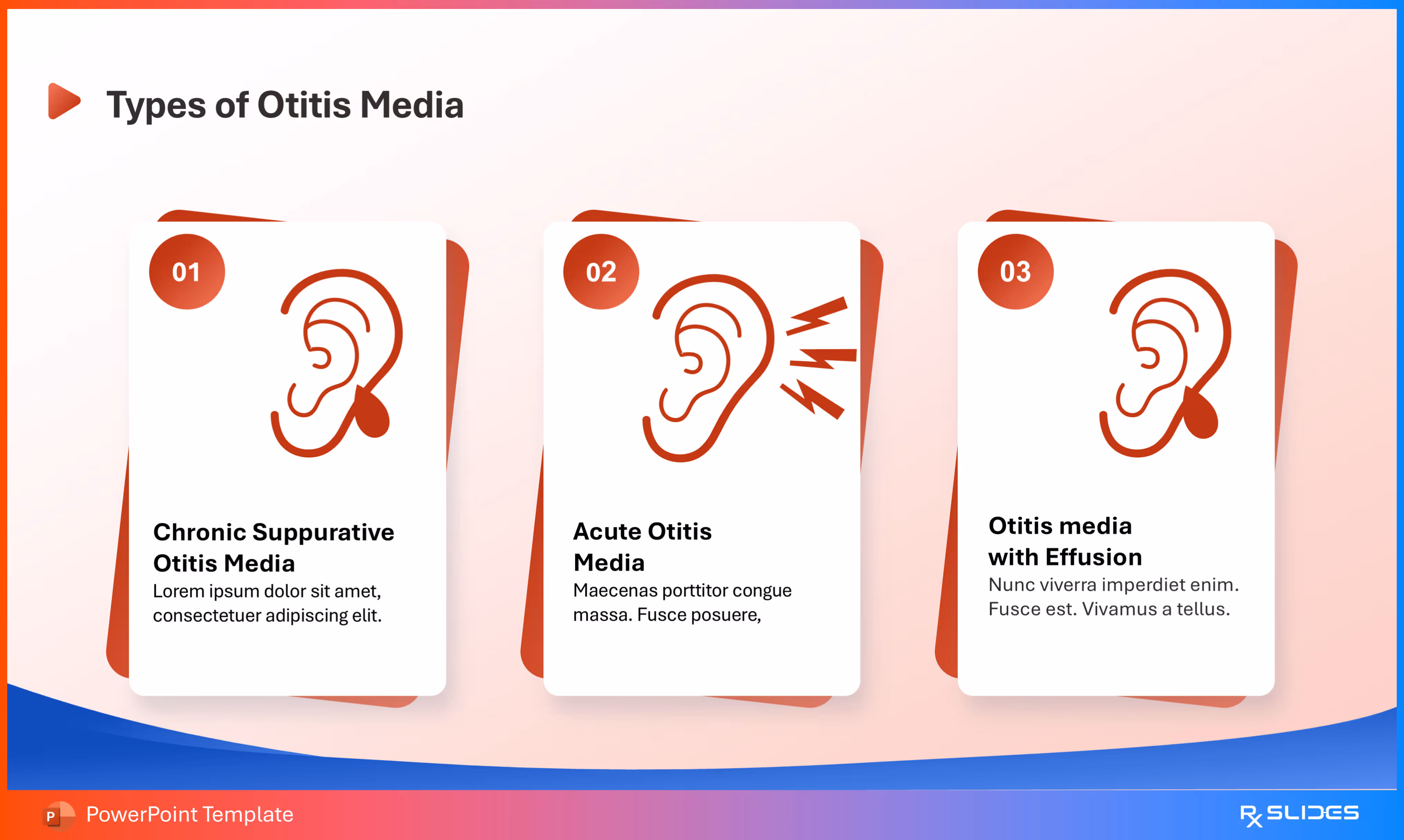
- Illustrates three primary types of Otitis Media.
- The design uses three vertical cards, numbered 01, 02, and 03, each containing a relevant ear icon and a description of the type: Chronic Suppurative Otitis Media, Acute Otitis Media, and Otitis media with Effusion.
Slide 24 - Acute Otitis Media (Key Symptoms)
.avif)
- Illustrates and explains the key clinical findings and symptoms specific to Acute Otitis Media (AOM).
- The slide features a large illustration of the ear pathology on the right, specifically showing the middle ear space filled with fluid draining outwards.
- Three key clinical features are highlighted in vertical, colorful boxes on the left: Pus in the Ear, Eardrum bulging, and Ear Drainage.
Slide 25 - Otitis Media With Effusion (OME)
.avif)
- Illustrates and explains the key clinical findings and consequences specific to Otitis Media with Effusion (OME).
- The slide features a large illustration of the ear pathology on the right, showing the middle ear space filled with fluid but without the intense inflammation seen in AOM.
- Two key clinical features are highlighted in vertical boxes on the left: Fluid accumulate and Hearing impaired.
Slide 26 - Otitis Media Symptoms (Section Title)
.avif)
- Section divider slide to transmit Symptoms of Otitis Media.
- The slide features a large, stylized blue and red thermometer icon, visually representing fever, a common symptom of acute infection.
- The title "Otitis Media Symptoms" is prominent on the left.
Slide 27 - Otitis Media Symptoms (Five Clinical Findings)
.avif)
- Illustrates five key symptoms associated with Otitis Media.
- The design uses a semi-circular arrangement of five content blocks surrounding a central title circle, creating an engaging visual flow: Heat, Loss of Appetite, Hearing Impairment, Pus from Ear, and a fifth point located at the top center with a thermometer icon.
Slide 28 - Otitis Media Symptoms (Detailed Breakdown)
.avif)
- Details four specific symptoms that patients with Otitis Media may experience, including neurological and general effects.
- The design uses a layout with four distinct content boxes, each containing an icon and a space for description: Hearing Impairment, Weakness, Dizziness, and Earache.
Slide 29 - Otitis Media Clinical Signs (Five Visual Findings)
.avif)
- Illustrates five clinical signs associated with Otitis Media.
- The design uses five vertical cards, each featuring a simplified illustration of a woman demonstrating the specific symptom: Hearing Impairment, Earache, Heat, Weakness, and Headache.
Slide 30 - Otitis Media Diagnosis (Section Title)
.avif)
- Section divider to transmit to the Diagnosis methods for Otitis Media.
- The slide features a large, stylized blue and red medical device icon.
- The title "Otitis Media Diagnosis" is prominent on the right.
- The design uses a soft pink, red, and blue color scheme.
Slide 31 - Diagnosis Of Otitis Media (Four Methods)
.avif)
- Illustrates four methods used in the clinical diagnosis of Otitis Media.
- The design uses a circular flow chart with four connected content blocks: Audiometry, Tympanometry, Medical history, and Physical examination.
Slide 32 - Otoscope Diagnosis (Physical Examination Tool)
.avif)
- Illustrates the role of the Otoscope in the physical examination component of Otitis Media diagnosis.
- The slide features a large illustration showing the external, middle, and inner ear, with an Otoscope inserted into the ear canal.
Slide 33 - Tympanometry Diagnosis (Assessment of Middle Ear)
.avif)
- Illustrates the role of Tympanometry in the diagnosis of Otitis Media.
- The slide features a large illustration showing the ear anatomy with a tuning fork near the ear canal.
- The illustration specifically highlights the middle ear and ossicles.
Slide 34 - Otitis Media Complications (Section Title)
.avif)
- Section divider slide to transmit to the Complications associated with Otitis Media.
- The slide features a large, stylized blue and red emergency siren/light icon.
- The title "Otitis Media Complications" is prominent on the left.
Slide 35 - Otitis Media Complications (Four Key Issues)
.avif)
- Illustrates four complications.
- The design uses a row of four diamond-shaped icons in contrasting red and blue, each with a brief description beneath: Infection spread, Hearing loss, Speech delay, and Ear Drum ruptures.
Slide 36 - Otitis Media Complications (Alternative Detailed View)
.avif)
- An alternative to Slide 35.
Slide 37 - Otitis Media Treatment (Section Title)
.avif)
- Section divider slide to transmit to Treatment options for Otitis Media.
- The slide features a large, stylized blue and red capsule/pill icon.
- The title "Otitis Media Treatment" is prominent on the right.
- The design uses a soft pink, red, and blue color scheme.
Slide 38 - Otitis Media Treatment (Five Key Strategies)
.avif)
- Details five key management strategies for treating Otitis Media.
- The design uses a visual list, where a central red circle containing the title is connected to five vertical cards, each describing a treatment method: Antibiotics, Pain relief, Observation, Ear drainage, and Managing Underlying conditions.
Slide 39 - Tympanostomy (Surgical Treatment)
.avif)
- Illustrates and explains the surgical procedure known as Tympanostomy.
- The slide features a large illustration showing the ear anatomy with a small tube inserted through the Tympanic Membrane into the middle ear space.
- The tube is labeled "Ear Tube" and is positioned to help drain the fluid and ventilate the middle ear, which is otherwise blocked by infection.
Slide 40 - Otitis Media Prevention (Section Title)
.avif)
- Section divider slide to transmit to Prevention strategies for Otitis Media.
- The slide features a large, stylized icon of two hands cradling a spine/bone structure.
- The title "Otitis Media Prevention" is prominent on the left.
Slide 41 - Otitis Media Prevention (Six Strategies)
.avif)
- Illustrates six strategies for preventing or reducing the risk of Otitis Media.
- The design uses a radial flow chart of six red circular icons surrounding a central blue ear icon, creating a comprehensive overview of protective measures: Stop colds, Chiropractic care, Manage stress, Eliminate allergens, Clean up your diet, and Breast feed.
Slide 42 - Thank You (Conclusion)
.avif)
- Concludes the presentation on Otitis Media.
- The slide features a large, central blue ear outline with red sound/pain lines radiating from it.
- The phrase "Thank You" is displayed in white text within a bold.
Features of the Template
- 100% editable PowerPoint template.
- Editable colors, you can change according to your presentation style and company branding guidelines.
Purchase Template
Purchase the fully editable PowerPoint open file template
Template Specifications
Slides count:
40+ Slides
Free monthly updates via email
Compatible with:
Microsoft PowerPoint
File type:
PPTX
Dimensions:
(16:9)