A Guide to Presenting Otitis Media with Our Medical Template
Otitis media is an inflammatory condition of the middle ear cavity and remains one of the most common pediatric diagnoses globally. Its pathophysiology centers on Eustachian tube dysfunction, middle ear effusion, and subsequent microbial proliferation. For medical educators and congress presenters, translating these mechanisms requires visuals that integrate anatomy, pathology, and clinical decision-making.
The Otitis Media PowerPoint Template provides a structured framework to teach this condition from foundational ear anatomy through diagnostic workflow and surgical intervention.
Below is a practical guide for leveraging the template in professional medical education settings.
1. Anatomical Orientation and Disease Localization
Begin by establishing normal ear structure before introducing pathology.
Otitis Media Anatomy
Use these visuals to outline the three anatomical regions:
Emphasize that otitis media develops posterior to the tympanic membrane within the middle ear cavity, creating the anatomical basis for effusion and pressure-related symptoms.
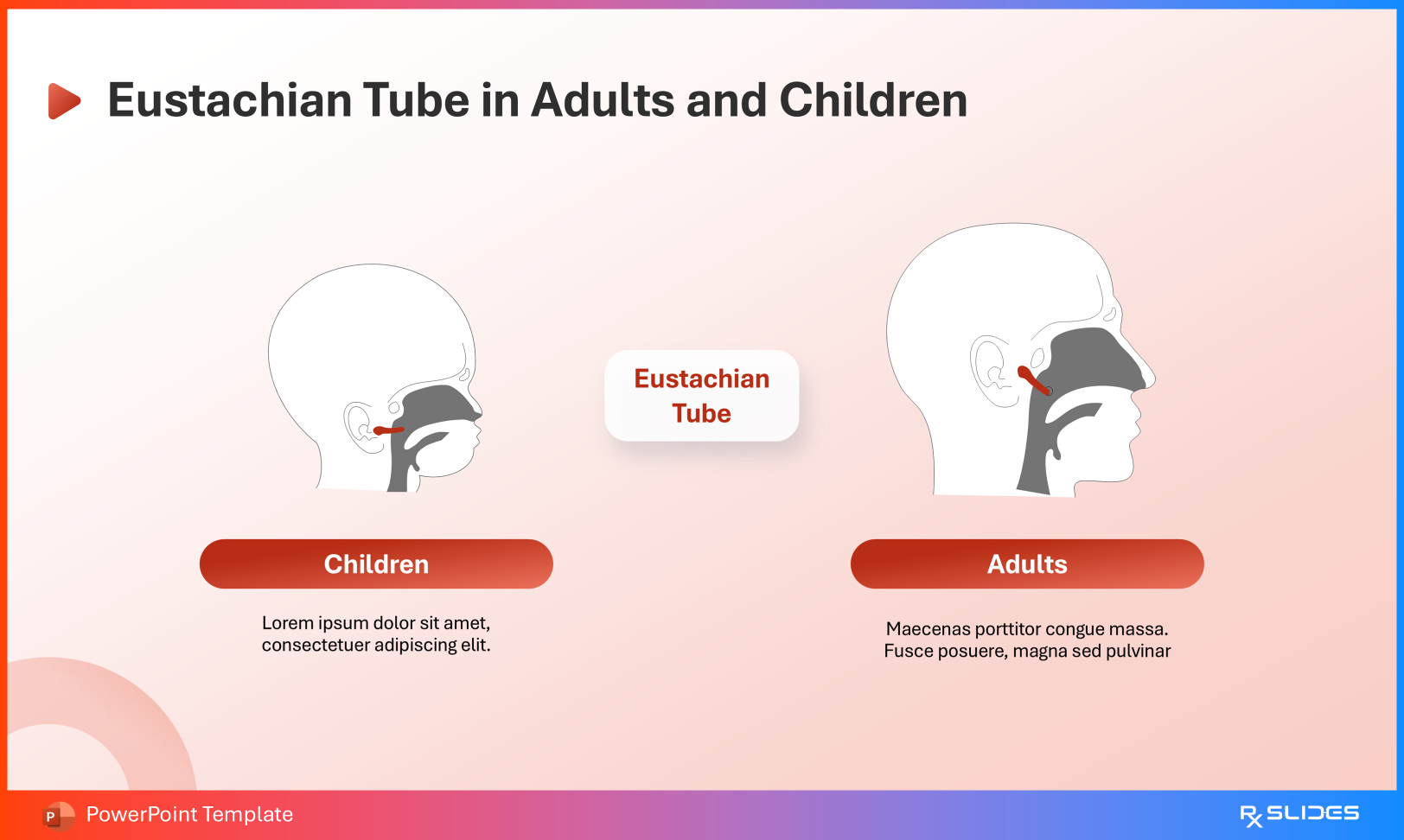
Eustachian Tube in Adults vs Children
This comparison is central to pediatric pathophysiology:
Pediatric Eustachian tubes are shorter, narrower, and more horizontal
This configuration compromises ventilation and drainage
Clinically, this explains the increased incidence of recurrent infections in infants and young children and supports anatomical reasoning during case-based teaching.
2. Disease Cascade: Pathogenesis for Medical Training
For pathology and microbiology education, present otitis media as a sequential process.
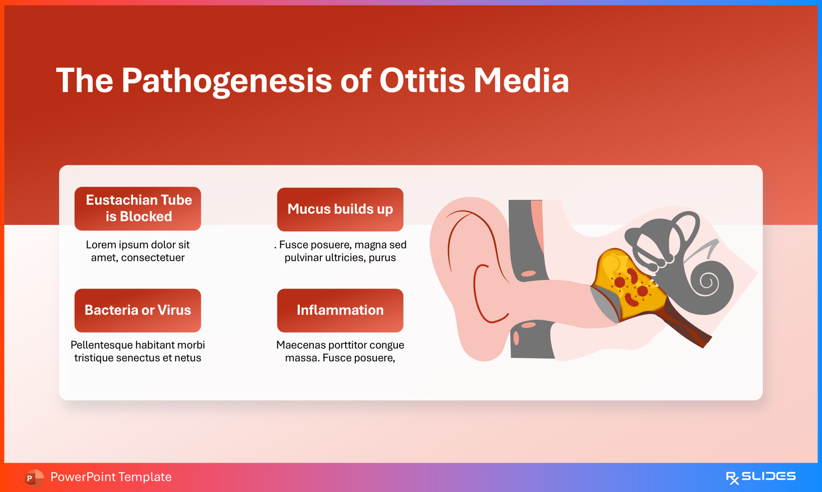
Pathogenesis of Otitis Media
Use the flow diagram to illustrate:
Eustachian tube obstruction, commonly following upper respiratory infection
Mucus retention within the middle ear
Bacterial or viral colonization
Local inflammatory response resulting in pain, pressure, and fever
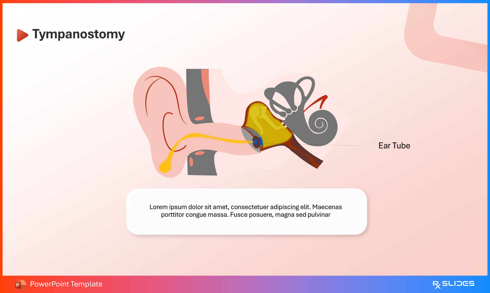
This template connects pediatric Eustachian tube anatomy with the clinical pathway from middle ear effusion to tympanostomy tube placement. It enables seamless progression from mucus accumulation and microbial proliferation to diagnostic otoscopy and long-term hearing preservation, supporting education across pediatrics, primary care training, and medical congress presentations.