A Guide to Presenting Scoliosis in PowerPoint

A Guide to Presenting Scoliosis in PowerPoint

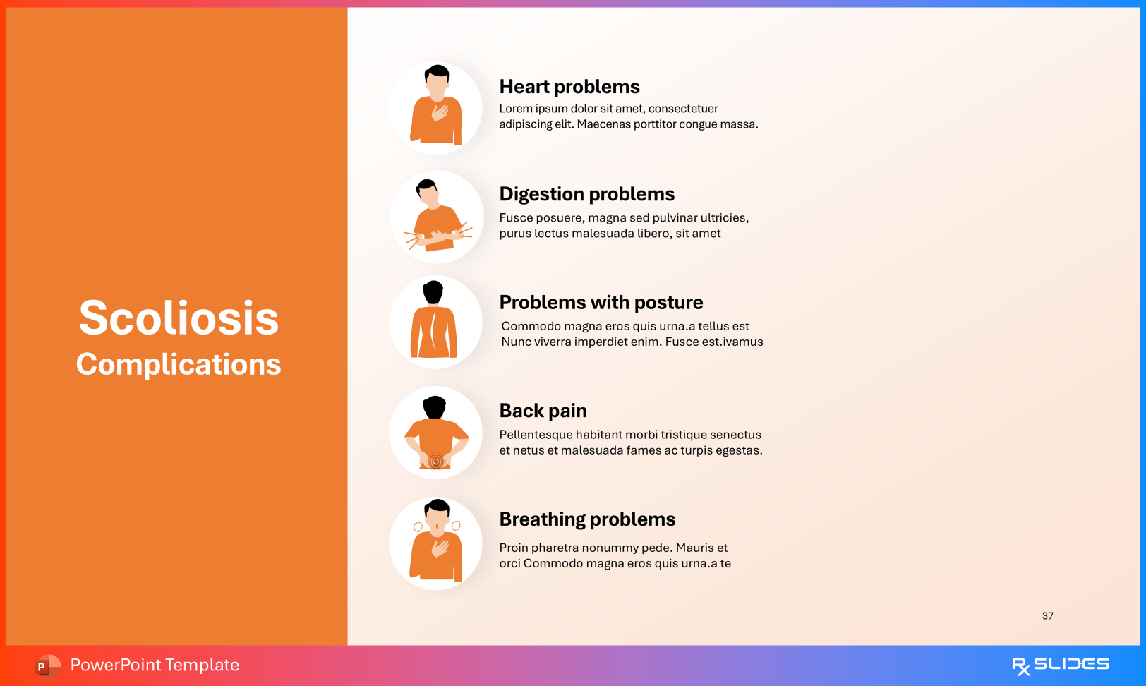
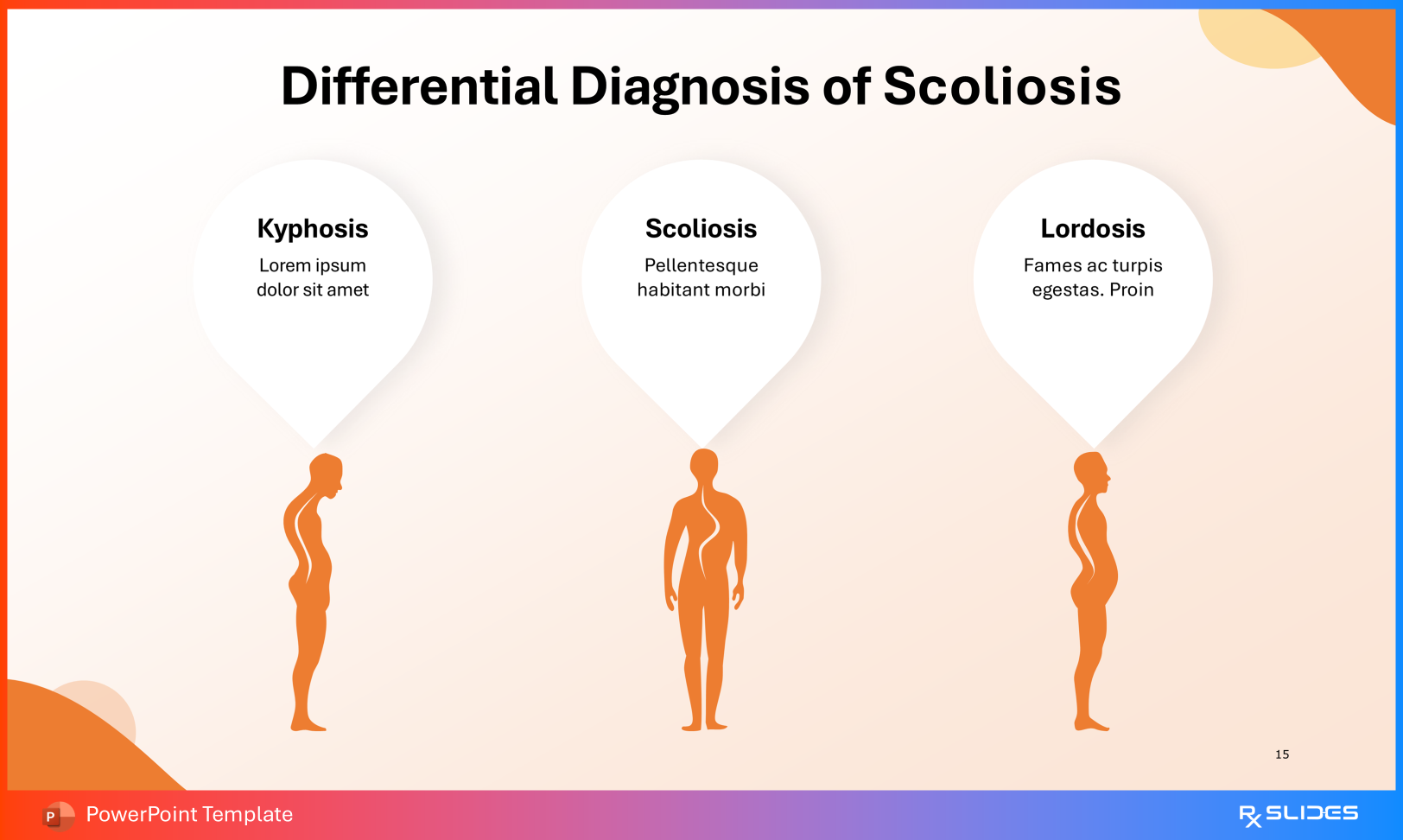
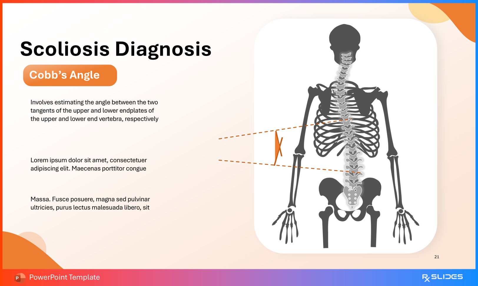
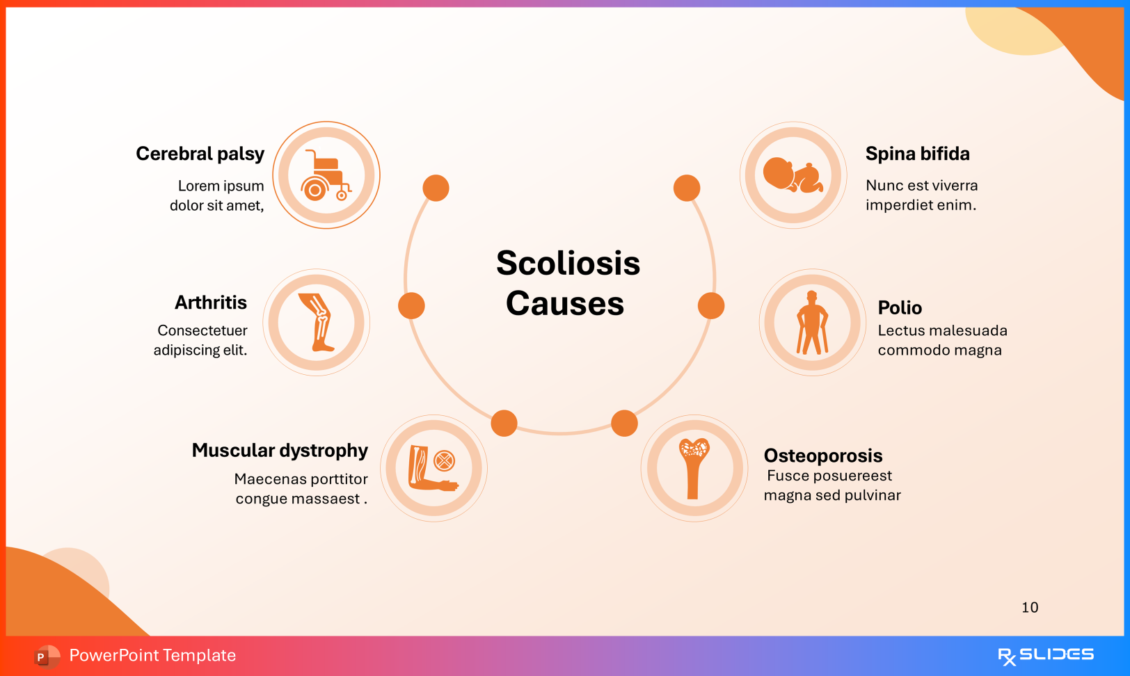
Scoliosis is a three-dimensional deformity of the spine characterized by lateral curvature accompanied by vertebral rotation. Explaining distinctions such as functional versus structural curves, or demonstrating the geometry of Cobb’s angle, requires precise visual support.

Whether addressing orthopedic residents, medical students, physical therapists, or patients and families, the Scoliosis PowerPoint Presentation Template provides a comprehensive framework for teaching spinal anatomy, diagnosis, and treatment pathways.

This guide outlines how to deploy each section effectively across clinical and educational settings.

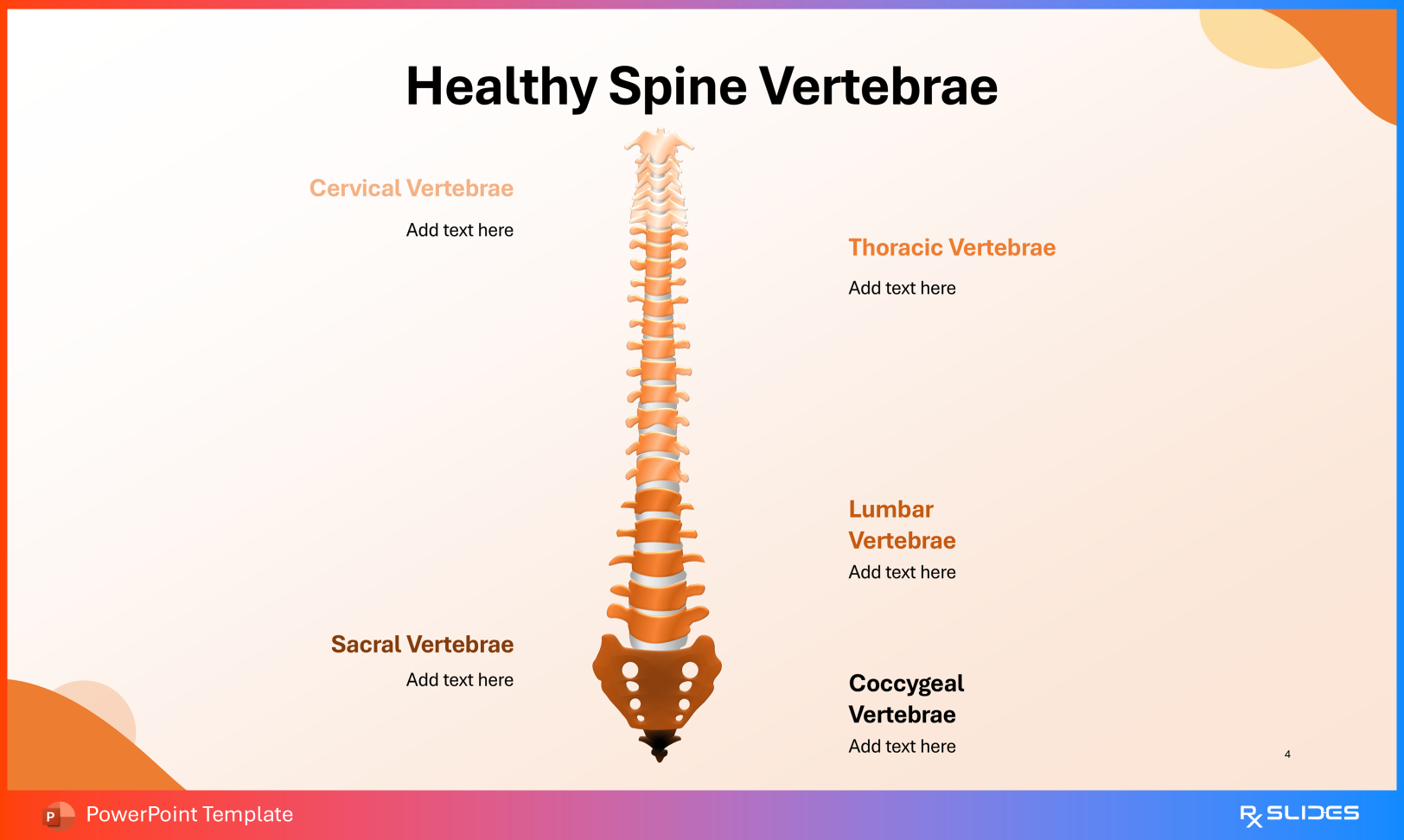
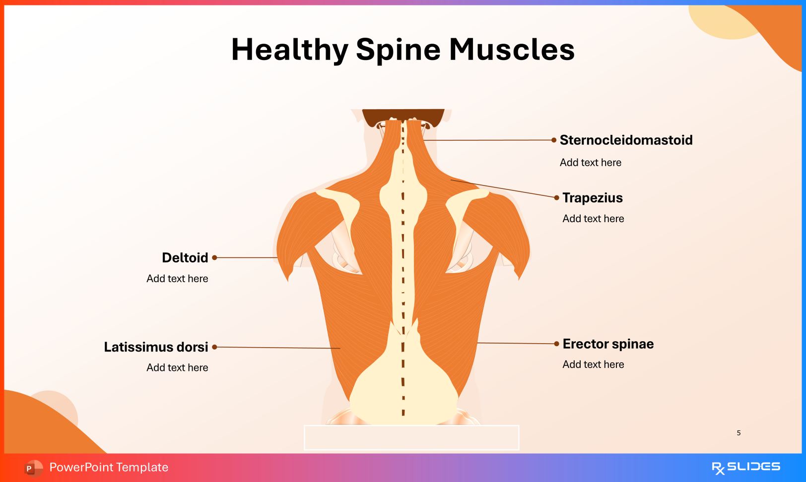
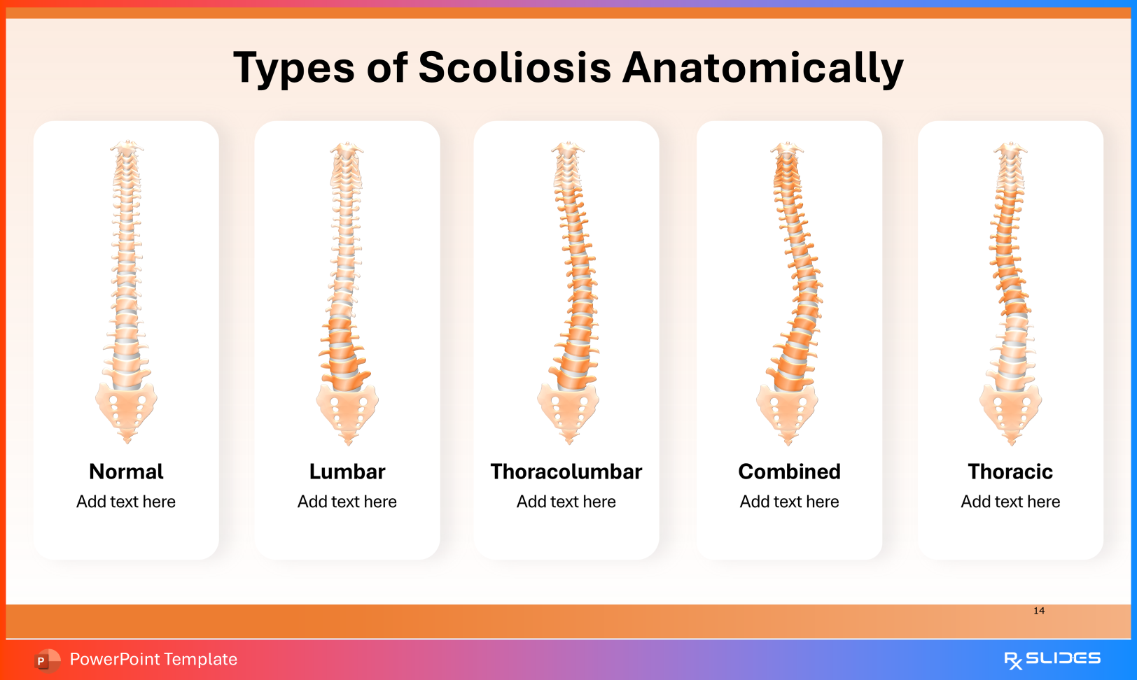
1. Establishing the Baseline: Normal Spine Anatomy

Before introducing pathology, learners must understand normal spinal alignment.

Healthy Spine Vertebrae

Use this slide to review regional anatomy:

  • Cervical
  • Thoracic
  • Lumbar
  • Sacral
  • Coccygeal
Healthy Spine Vertebrae Slide from Scoliosis PowerPoint Presentation Template

This provides the anatomical reference point for identifying abnormal curvature.

Healthy Spine Muscles

Highlight stabilizing structures including:

  • Erector spinae
  • Latissimus dorsi
  • Trapezius
Healthy Spine Muscles from Scoliosis PowerPoint Presentation Template

This contextualizes how muscular imbalance and asymmetrical loading may contribute to curve progression.

2. Clinical Classification and Differential Diagnosis

Diagnosis extends beyond visual inspection and requires anatomical categorization.

Anatomical Types of Scoliosis

Use these visuals to classify curves as:

  • Thoracic
  • Lumbar
  • Thoracolumbar
  • Combined
Types of Scoliosis Anatomically Slide from Scoliosis PowerPoint Presentation Template

This is essential for treatment planning and prognostication.

Differential Diagnosis

Contrast scoliosis with other sagittal plane deformities:

  • Kyphosis (excessive posterior curvature)
  • Lordosis (exaggerated anterior curvature)
Differential Diagnosis Slide from Scoliosis PowerPoint Presentation Template

These comparisons reinforce the coronal-plane nature of scoliosis.

3. Quantifying Severity: Cobb Angle Measurement

Cobb angle remains the clinical gold standard for curve assessment.

Cobb’s Angle Indications

Use this slide to demonstrate:

  • Drawing lines along the superior endplate of the upper end vertebra and inferior endplate of the lower end vertebra
  • Measuring the intersecting angle
Cobb’s Angle Indications Slide from Scoliosis PowerPoint Presentation Template

Severity classification:

  • 10°–20°: Mild scoliosis
  • 20°–40°: Moderate scoliosis

40°: Severe scoliosis

Cobb’s Angle Indications Slide from Scoliosis PowerPoint Presentation Template

This section is central for monitoring progression and determining intervention thresholds.

4. Patient-Centered Education: Symptoms and Etiology

For patients and caregivers, emphasis shifts from measurements to observable changes.

Scoliosis Symptoms

Use graphics to illustrate common findings:

  • Head not centered over pelvis
  • Asymmetric shoulder blades
  • Uneven hips or shoulders
Scoliosis Symptoms Slide from Scoliosis PowerPoint Presentation Template

These visuals validate patient concerns and improve engagement.

Causes of Scoliosis

Explain etiological categories:

  • Idiopathic (most common)
  • Congenital
  • Neuromuscular
Causes of Scoliosis Slide from Scoliosis PowerPoint Presentation Template

The template supports discussion of associated conditions such as cerebral palsy, muscular dystrophy, spina bifida, and poliomyelitis.

5. Management Spectrum: Conservative Care to Surgical Correction

Treatment depends on age, skeletal maturity, curve magnitude, and progression.

Conservative Treatment

Outline nonoperative strategies:

  • Observation
  • Bracing
  • Physical therapy and exercise
  • Analgesics or spinal injections for symptom control
Conservative Treatment Slide from Scoliosis PowerPoint Presentation Template

Posterior Spinal Fusion

For severe or progressive curves, use surgical visuals to explain:

  • Posterior exposure of the spine
  • Placement of pedicle screws and rods for deformity correction
  • Use of autograft bone (often from rib or iliac crest) to promote fusion
Posterior Spinal Fusion Slides from Scoliosis PowerPoint Presentation Template

This stepwise depiction supports informed consent and resident education.

6. Long-Term Perspective: Complications of Untreated Severe Scoliosis

Use the complications slides to explain potential consequences of advanced deformity:

  • Restrictive lung disease
  • Cardiac compromise
  • Gastrointestinal compression
Complications of Scoliosis Slide from Scoliosis PowerPoint Presentation Template

This reinforces the importance of timely monitoring and intervention.

Conclusion: From Cobb Angle Geometry to Functional Outcomes

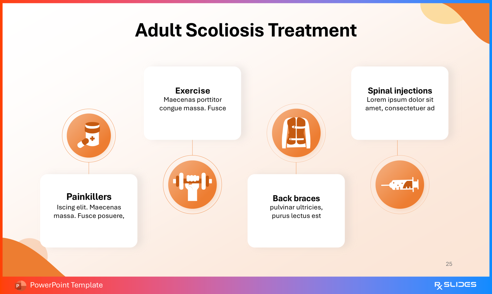
This template bridges quantitative assessment with real-world clinical impact. It allows educators to move fluidly from spinal biomechanics to brace management, and from neuromuscular etiologies to complex fusion surgery.

For healthcare professionals, it provides a cohesive visual narrative that supports anatomy instruction, clinical decision-making, patient counseling, and multidisciplinary care.

Latest Articles

Explore All