Urinary incontinence represents a prevalent lower urinary tract disorder with multifactorial etiologies spanning anatomical disruption, neuromuscular dysfunction, and age-related connective tissue changes. Despite its frequency, effective education requires clear visualization of both peripheral structures and central neural pathways governing micturition.
For urologists, neurologists, pelvic floor specialists, and medical educators, communicating these mechanisms demands tools that integrate anatomy, physiology, and clinical workflow.
The Urinary Incontinence PowerPoint Presentation Template provides a structured visual framework to support this integrated teaching approach.
Below is a guide for leveraging the template in professional medical education.
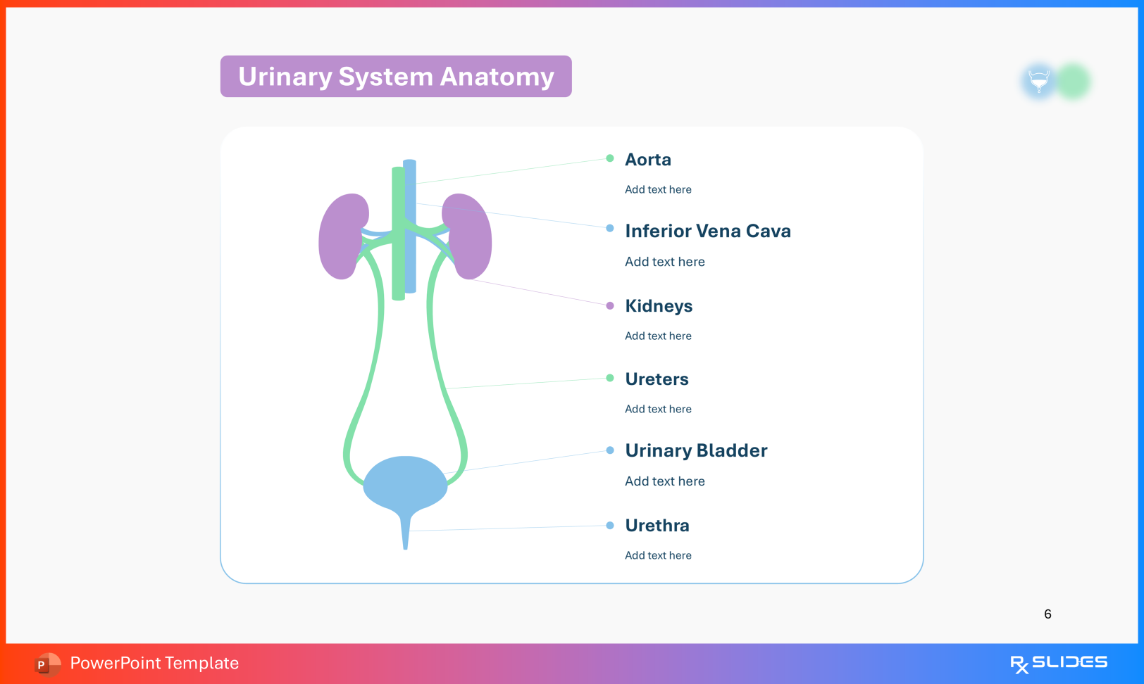
1. Lower Urinary Tract Anatomy: Structural Foundations of Continence Accurate diagnosis and management begin with anatomical clarity.
Urinary System Anatomy Urinary System Anatomy Slide from Urinary Incontinence PowerPoint Presentation Template Use this slide to trace urinary flow from the kidneys through the ureters into the bladder and urethra, establishing the macroscopic pathway.
Urinary Bladder Anatomy Urinary Bladder Anatomy Slide from Urinary Incontinence PowerPoint Presentation Template Highlight key continence structures:
Detrusor muscle Internal urethral sphincter External urethral sphincter These components illustrate coordinated storage and voiding mechanics.
Male and Female Anatomy Female Urinary System Anatomy Slide From Urinary Incontinence PowerPoint Presentation Template Male Urinary System Anatomy Slide from Urinary Incontinence PowerPoint Presentation Template Contrast sex-specific anatomy by demonstrating:
Prostatic positioning relative to the bladder outlet in males Pelvic organ relationships in females, including reproductive structures and bladder support This comparison supports understanding of sex-dependent incontinence mechanisms.
2. Neural Regulation of Micturition For neurology trainees and residents, bladder control must be framed as a centrally regulated reflex.
Neural Supply of Micturition Use this diagram to illustrate connections between:
Cerebral cortex Pontine micturition center Spinal cord pathways Pelvic and pudendal nerves Neural Supply of Micturition Slide from Urinary Incontinence PowerPoint Presentation Template This network governs voluntary and involuntary bladder activity.
Micturition Reflex Animation Walk through the physiologic sequence:
Bladder stretch receptors transmit afferent signals to the spinal cord and brain Descending inhibitory input maintains sphincter closure during storage Removal of inhibition initiates coordinated detrusor contraction and sphincter relaxation Micturition Reflex Animation Slide from Urinary Incontinence PowerPoint Presentation Template These visuals clarify neurogenic contributions to incontinence.
3. Clinical Classification and Diagnostic Strategy Precise phenotyping guides therapeutic selection.
Types of Urinary Incontinence Differentiate:
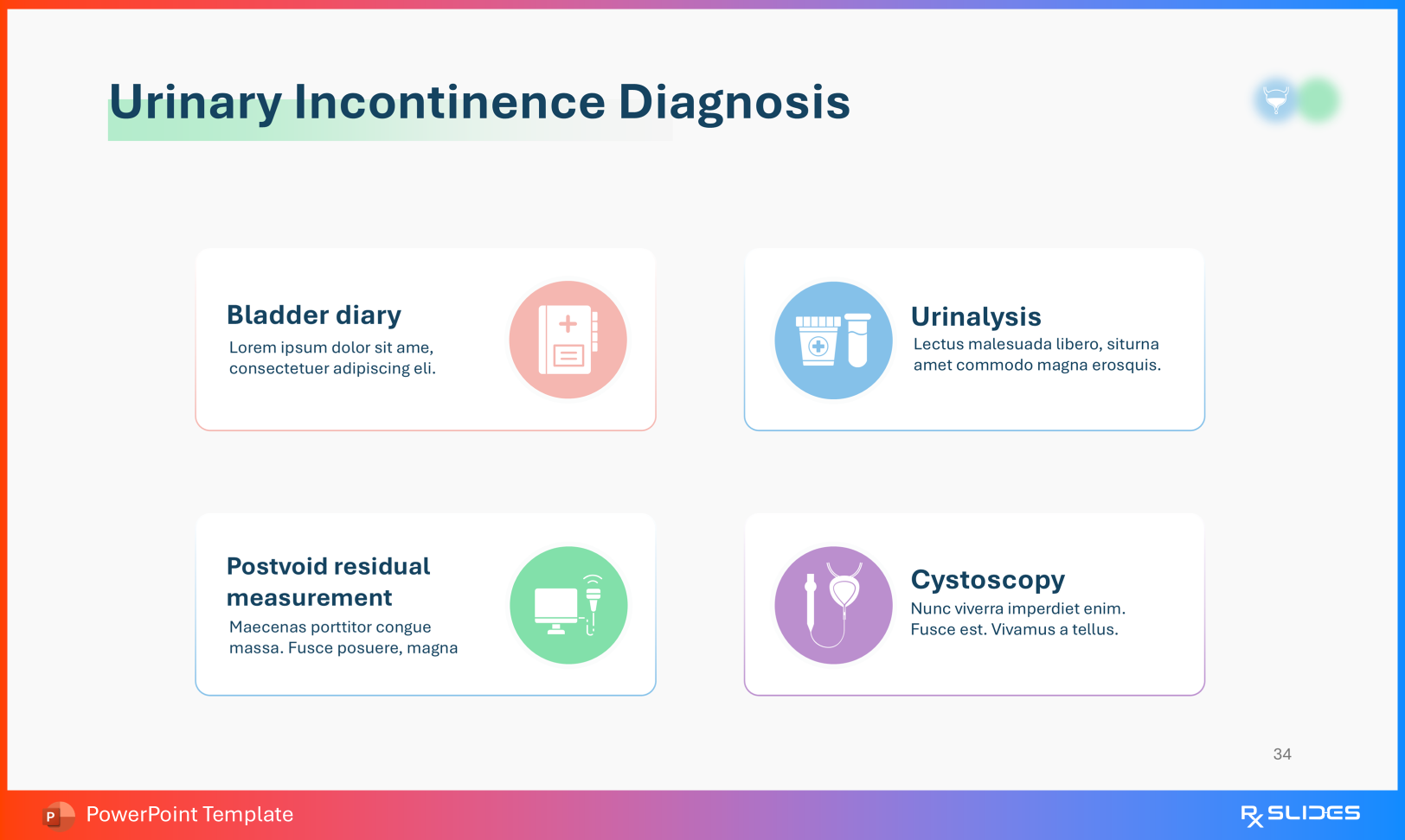
Stress incontinence: leakage with increased intra-abdominal pressure Urge incontinence: involuntary loss preceded by urgency Overflow incontinence: chronic retention with continuous dribbling Urinary Incontinence Types Slide from Urinary Incontinence PowerPoint Presentation Template Diagnostic Workflow Use the diagnosis and differential slides to outline evaluation tools:
Urinalysis Postvoid residual measurement Cystoscopy Urinary Incontinence Diagnosis Slide from Urinary Incontinence PowerPoint Presentation Template Simultaneously exclude alternative etiologies such as bladder stones or malignancy.
4. Risk Factors, Etiology, and Preventive Strategies For multidisciplinary education, understanding contributing factors supports early intervention.
Risk Factors Review variables including:
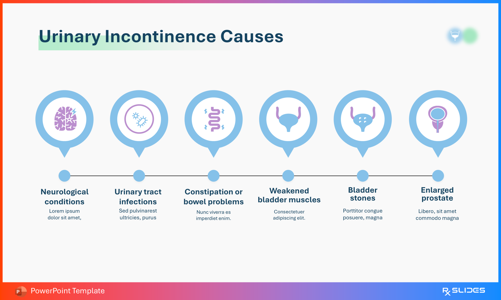
Advanced age Obesity Pregnancy and childbirth Genetic predisposition Urinary Incontinence Risk Factors Slide from Urinary Incontinence PowerPoint Presentation Template Causes Explain mechanistic contributors such as:
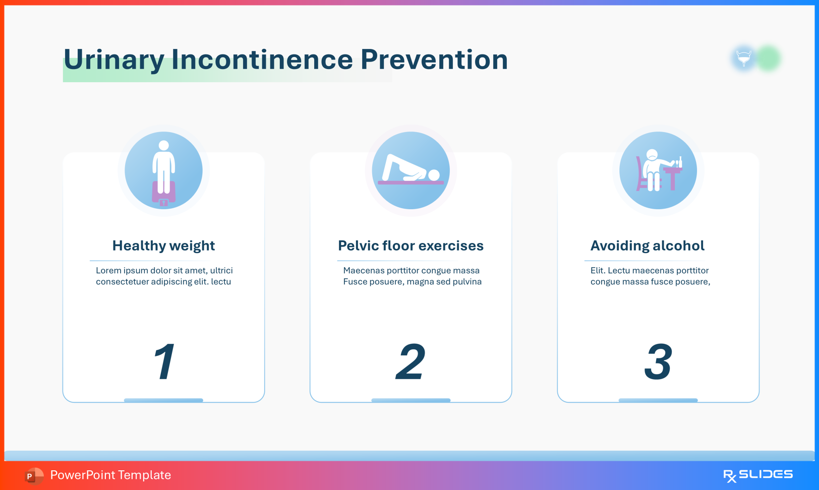
Detrusor instability Pelvic floor weakness Constipation Prostatic enlargement Urinary Incontinence Causes Slide from Urinary Incontinence PowerPoint Presentation Template Prevention and Treatment Demonstrate conservative and medical management options:
Pelvic floor muscle training Weight optimization Behavioral bladder training Pharmacologic therapy Surgical intervention when indicated Urinary Incontinence Treatment Slide from Urinary Incontinence PowerPoint Presentation Template Urinary Incontinence Prevention Slide from Urinary Incontinence PowerPoint Presentation Template These slides support evidence-based care pathways.
Conclusion: From Signaling to Pelvic Floor Rehabilitation This template bridges central neural regulation with peripheral anatomical function, allowing educators to move seamlessly from pontine micturition pathways to clinical strategies such as pelvic floor strengthening and surgical correction.
For healthcare professionals, it offers a coherent framework to teach continence physiology, diagnostic reasoning, and therapeutic management across urology, neurology, geriatrics, and pelvic health.