Astigmatism Educational Presentation Framework Using a Structured Medical Slide Template
Explaining refractive errors to a diverse audience whether they are anxious patients, medical students, or hospital administrators requires high-quality visuals and a structured narrative. The Astigmatism PowerPoint Presentation Template provides a comprehensive toolkit to demystify this common eye condition.
Here is a guide on how to leverage the specific features of this template to create an engaging and educational presentation.
1. Visualizing the Anatomy: Setting the Stage
Before explaining what goes wrong, you must explain how the eye is supposed to work. The template offers detailed Eye Anatomy slides that are essential for any audience level.
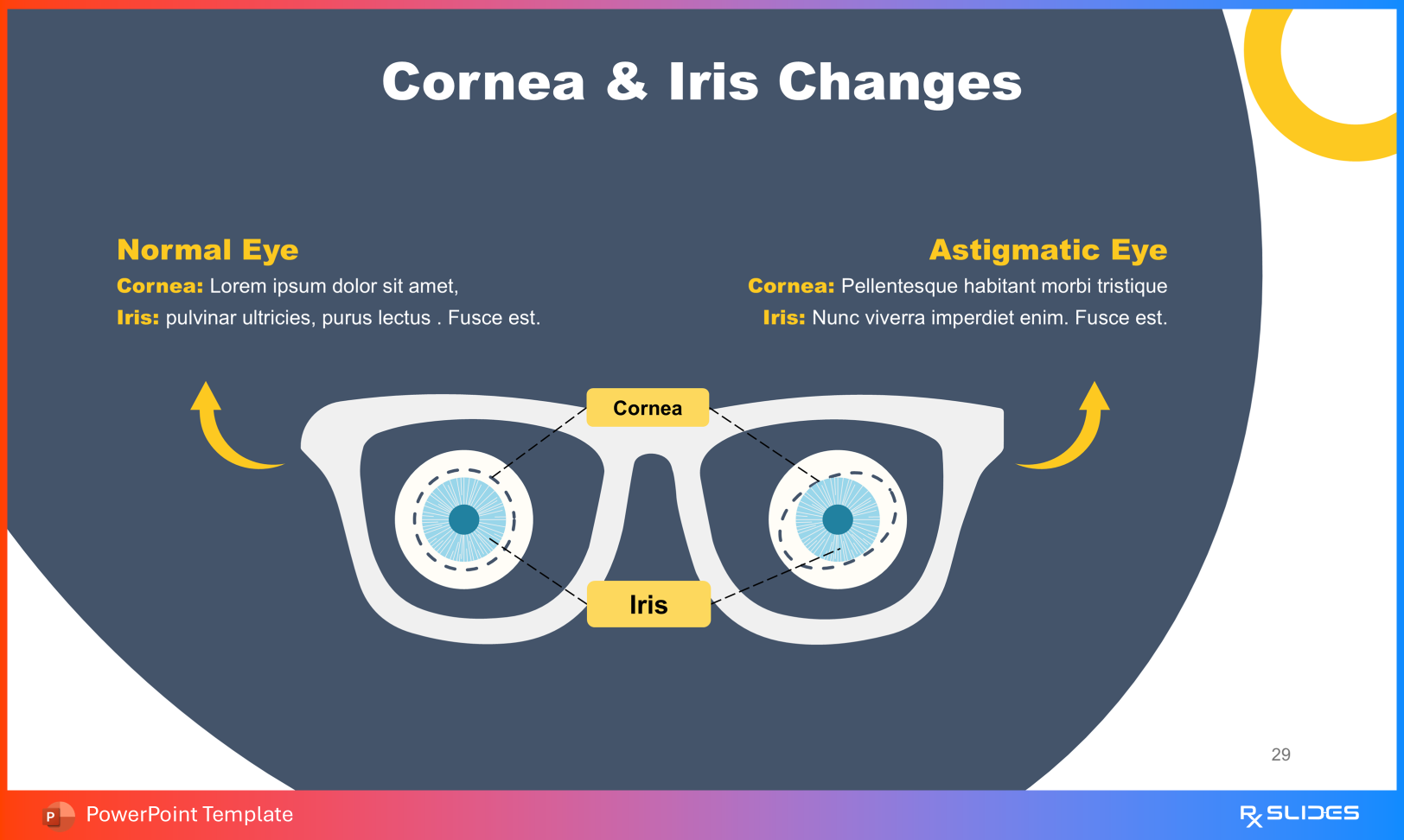
Use the "Anatomy of the eye" slide to point out the Cornea and Lens, explaining that these are the "windows" light must pass through.
You can delve deeper using the labeled diagrams of the Retina Structure, Tear Film Components (Lipid, Aqueous, and Mucin layers), and Glandular Structures like the Lacrimal and Meibomian glands.
2. The Mechanics of Blur: Refraction and Pathophysiology
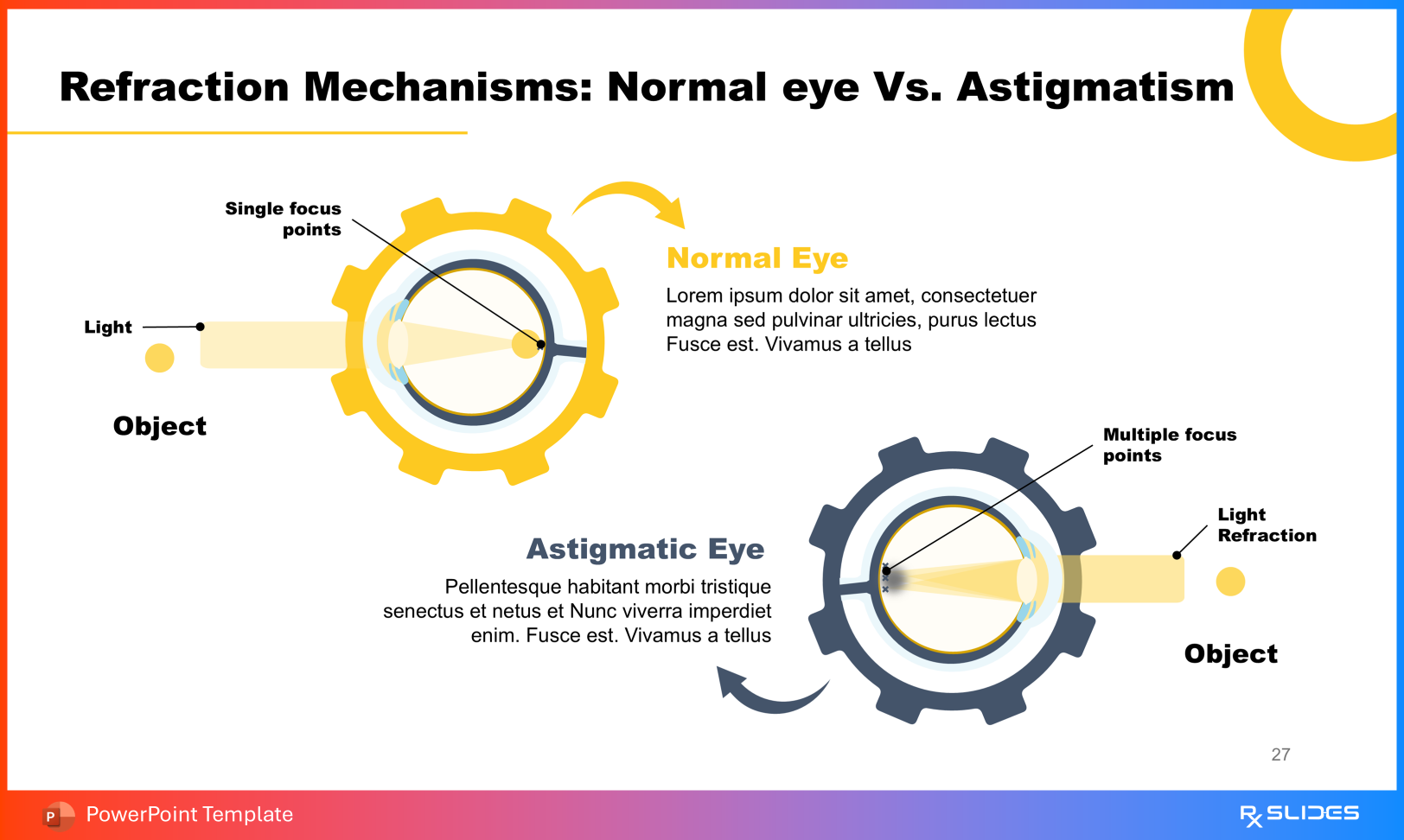
The most complex concept to convey is how the shape of the eye distorts light. The template excels here by offering side-by-side comparisons of a "Normal Eye" versus an "Astigmatic Eye".
Example Slide: Refraction Mechanisms
This slide visually demonstrates how light enters the eye. It contrasts the single focus point of a normal eye against the multiple focus points found in astigmatism, which causes the blurred vision.
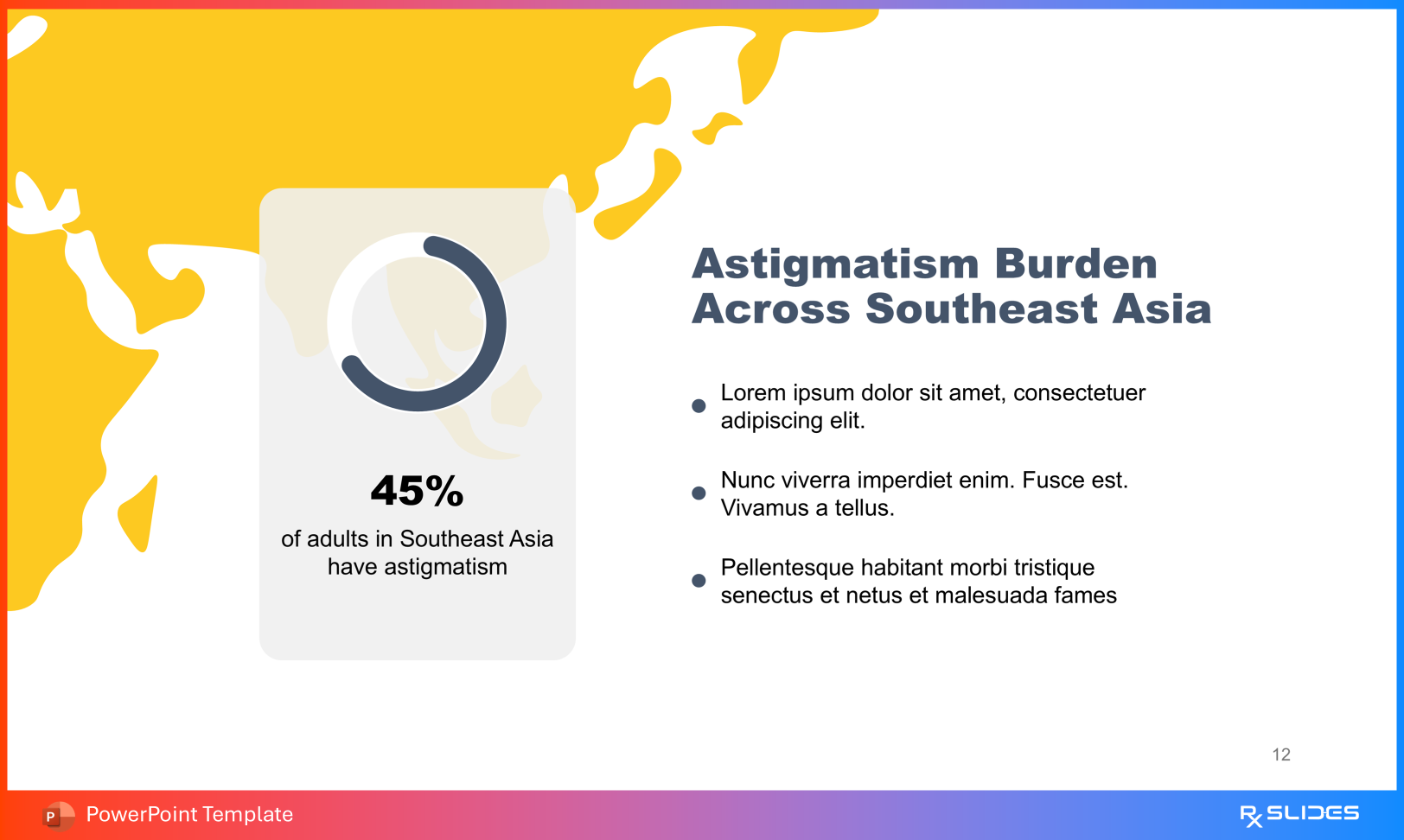
If you are presenting to public health officials or conducting an epidemiological review, the template provides robust data visualization tools.
Prevalence: Use the "Astigmatism Around the World" and "Astigmatism Rates Across Selected Countries" slides. These include map graphics and percentage placeholders (e.g., "Xx% of adults in Southeast Asia") to display regional data effectively.
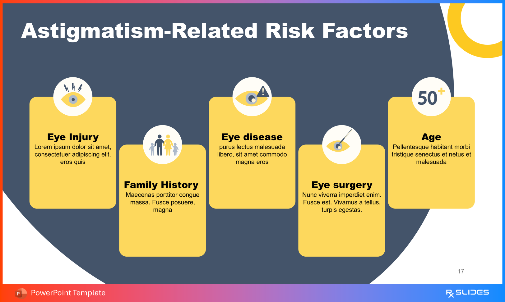
Risks: To discuss etiology, utilize the "Underlying Risk Contributors" slide. It features icons representing key factors such as Eye Injury, Family History, Eye Disease, Eye Surgery, and Age, making it easy to memorize or list during a lecture.
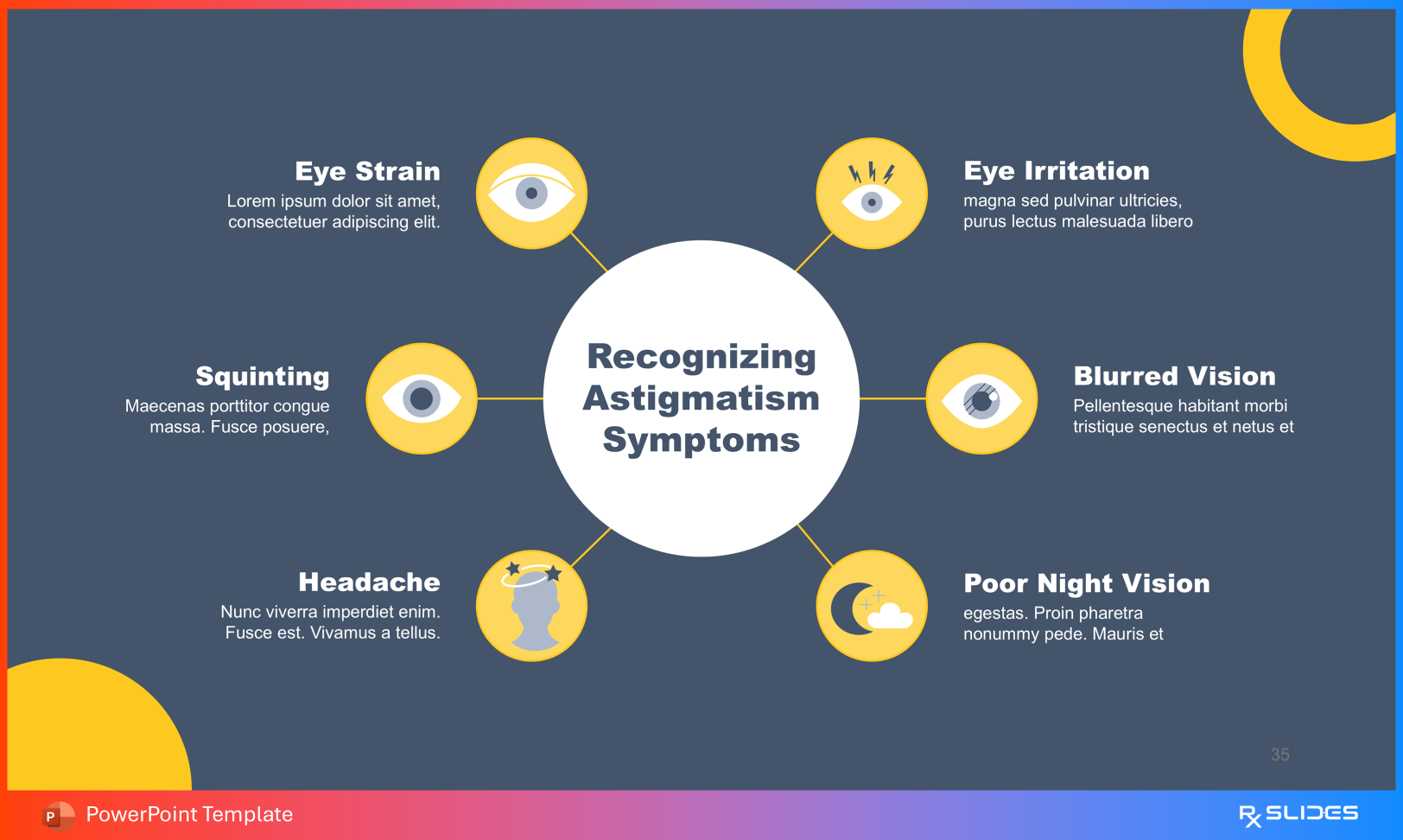
When training healthcare staff or educating patients on warning signs, the Symptoms and Diagnosis sections are critical.
Identifying Symptoms: The template uses intuitive icons for Eye Strain, Headache, Eye Irritation, Squinting, and Poor Night Vision. This helps patients quickly recognize their own experiences on the screen.
Diagnostic Tools: You can walk an audience through the examination process using slides dedicated to Pupil Dilatation, Refraction tests, and Visual Acuity Tests.
The template moves beyond problems to offer concrete solutions, ranging from non-invasive options to surgical interventions.
Example Slide: Laser Vision Correction This is a standout feature for surgical consultations. It breaks down the procedure into four distinct, illustrated steps: Eye Drop Anesthesia, Corneal Flap Creation, Excimer Laser Irradiation, Return The Flap.
Finally, wrap up your presentation with actionable advice using the "Astigmatism Prevention Tips" slide. This section allows you to highlight lifestyle habits such as Regular Eye Exams, Eye Hygiene, and proper Eye Protection.
This template transforms abstract optical concepts into clear, visual stories. It allows you to take your audience on a journey from the cellular structure of the retina to the global prevalence of the condition, and finally to the step-by-step mechanics of laser surgery.