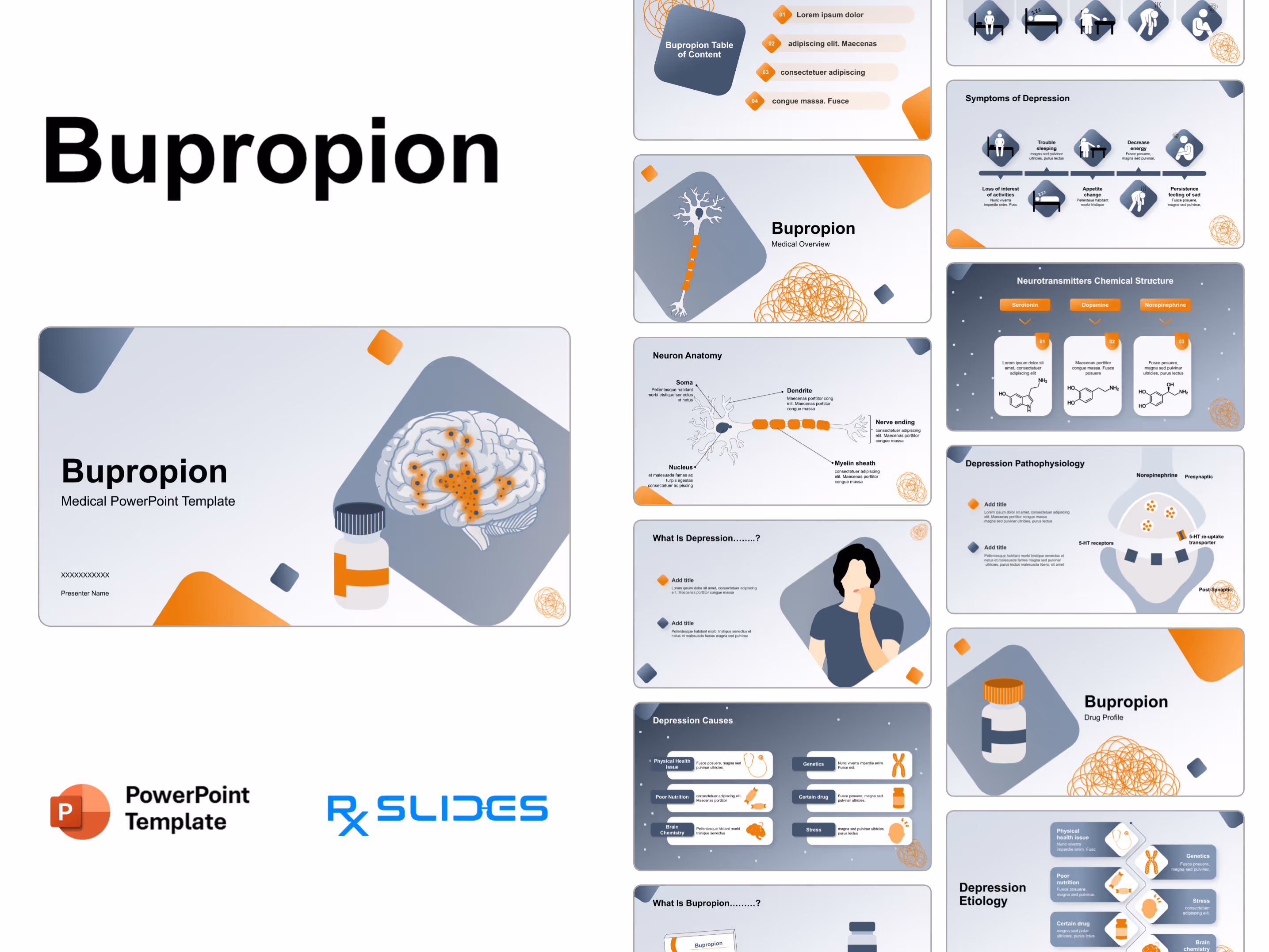
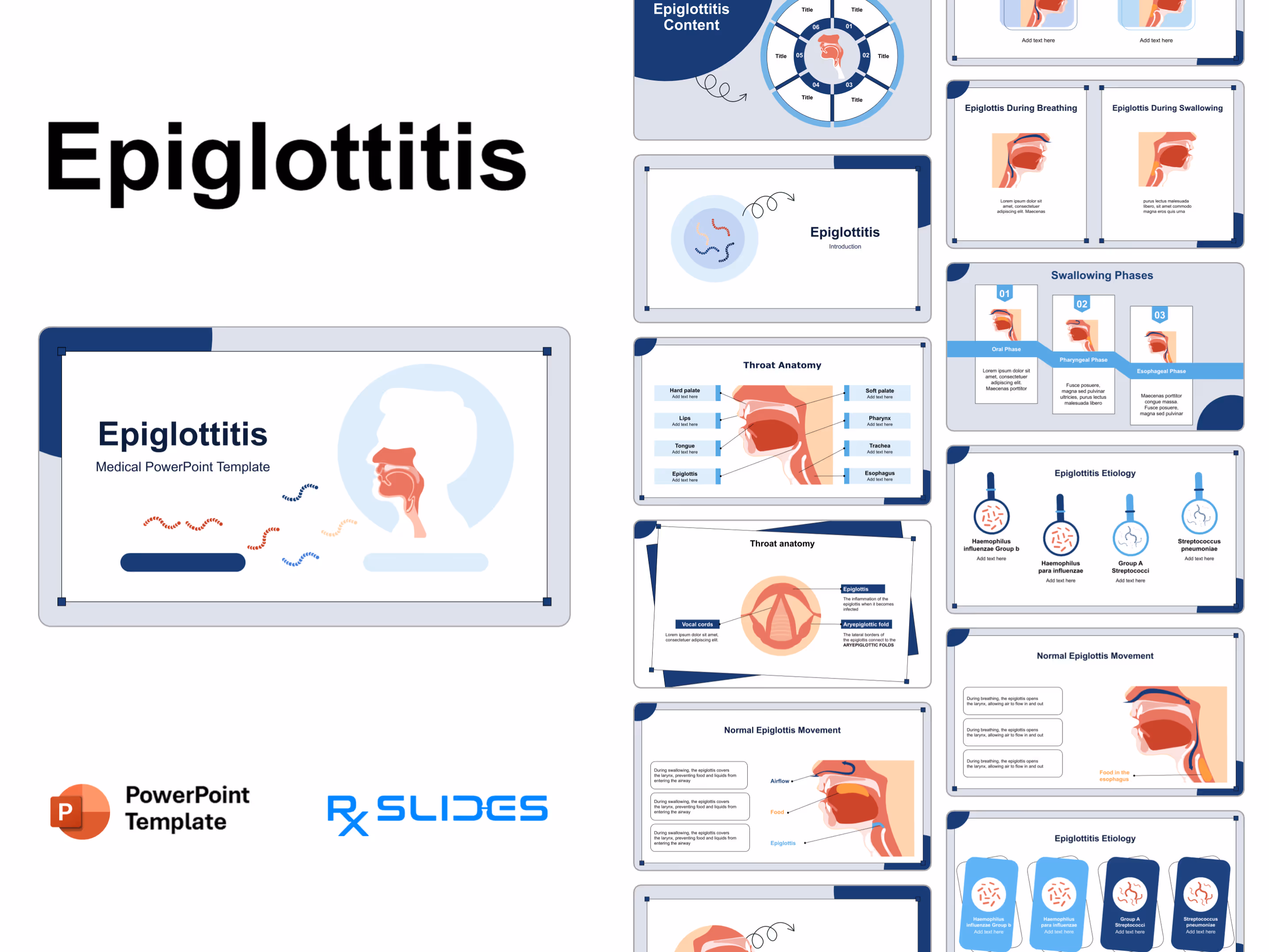
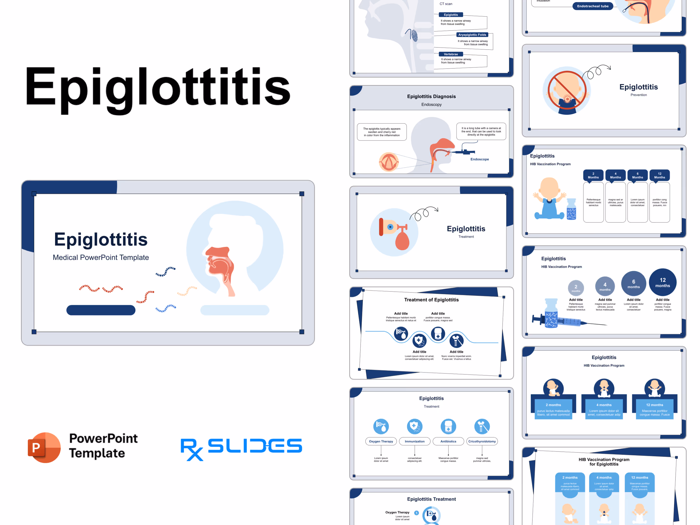
Epiglottitis PowerPoint Template
No items found.
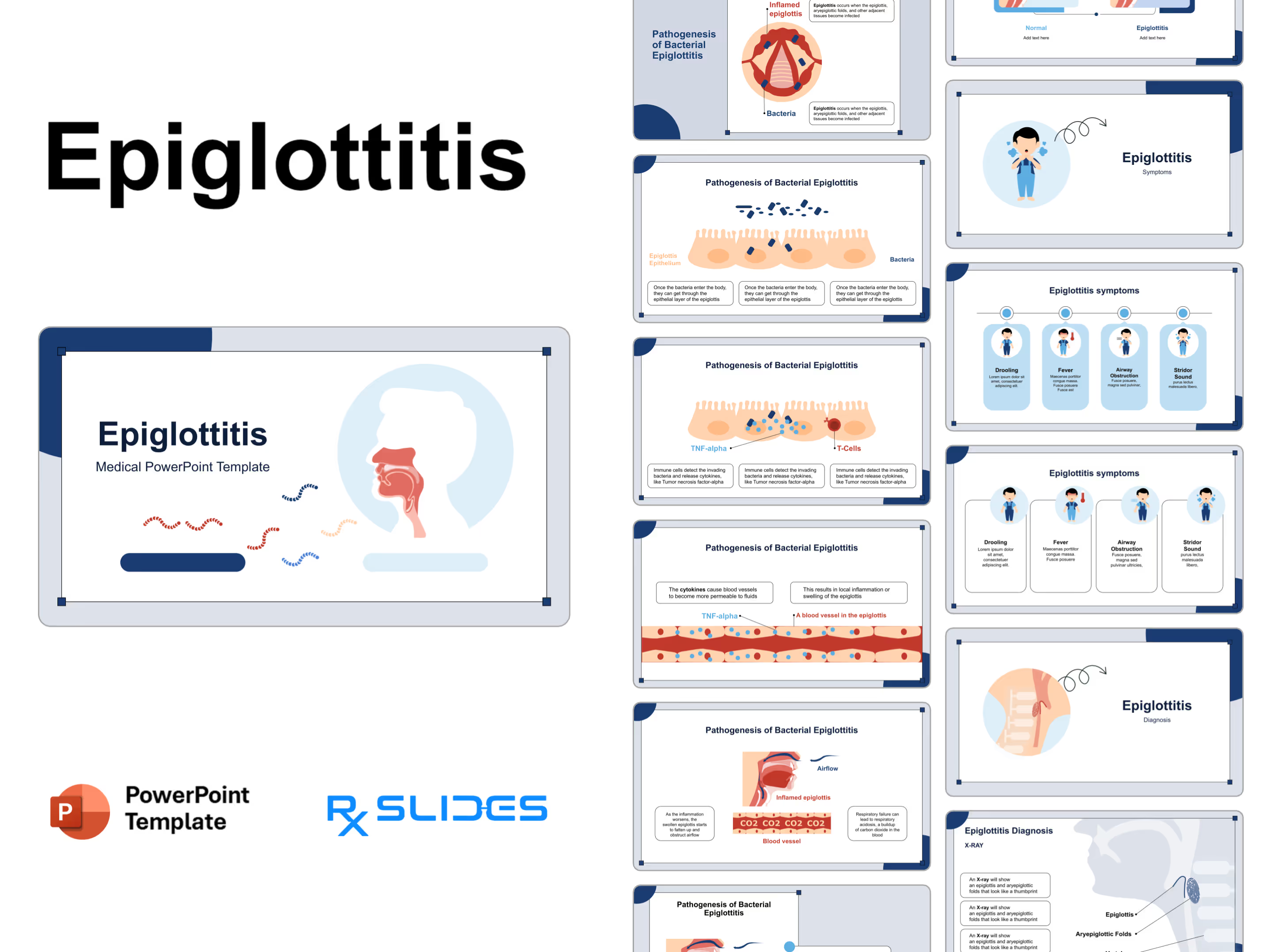
The Epiglottitis Presentation: Medical PowerPoint Template
- The Epiglottitis PPT template is an animated medical PowerPoint template that will help you realize the full potential of your presentation.
- Our carefully designed slides, which include medical animations and infographics, will attract your audience.
- You can rely on our demonstrated infographics to provide a dynamic and appealing Epiglottitis presentation to your audience.
- Go beyond basic Respiratory Diagrams with our interactive Respiratory System Templates, which allow you to represent more profoundly, promoting deeper engagement and understanding.
Epiglottitis PowerPoint Template Preview
Epiglottitis PowerPoint Template Content
Slide 1 - Epiglottitis Introduction (Title Slide)
.avif)
- introduces the presentation on Epiglottitis with a professional and engaging medical design.
- The slide prominently features a simplified anatomical cross-section of the head and throat.
Slide 2 - Epiglottitis Table of Contents (Navigation)
.avif)
- organizes the presentation and clearly outline six main discussion points for your audience.
- The design features a central graphic of the affected throat area surrounded by six numbered content sections.
Slide 3 - Introduction Section Divider
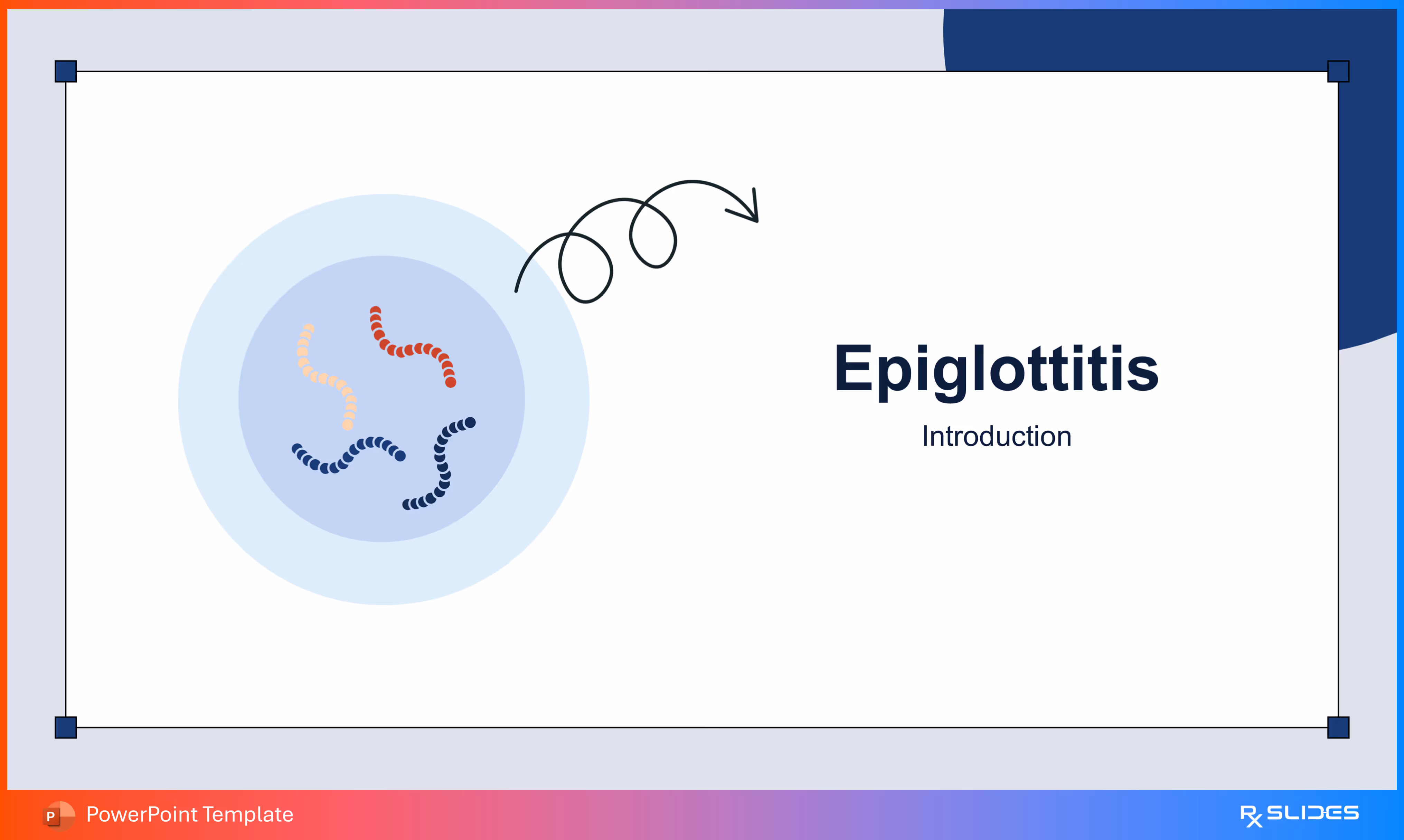
- divider slide to clearly signal the start of the Introduction section for the Epiglottitis presentation.
- The design features stylized, curved bacterial shapes in the corner, visually representing the infectious nature of the medical topic without relying on complex diagrams.
Slide 4 - Throat Anatomy Diagram
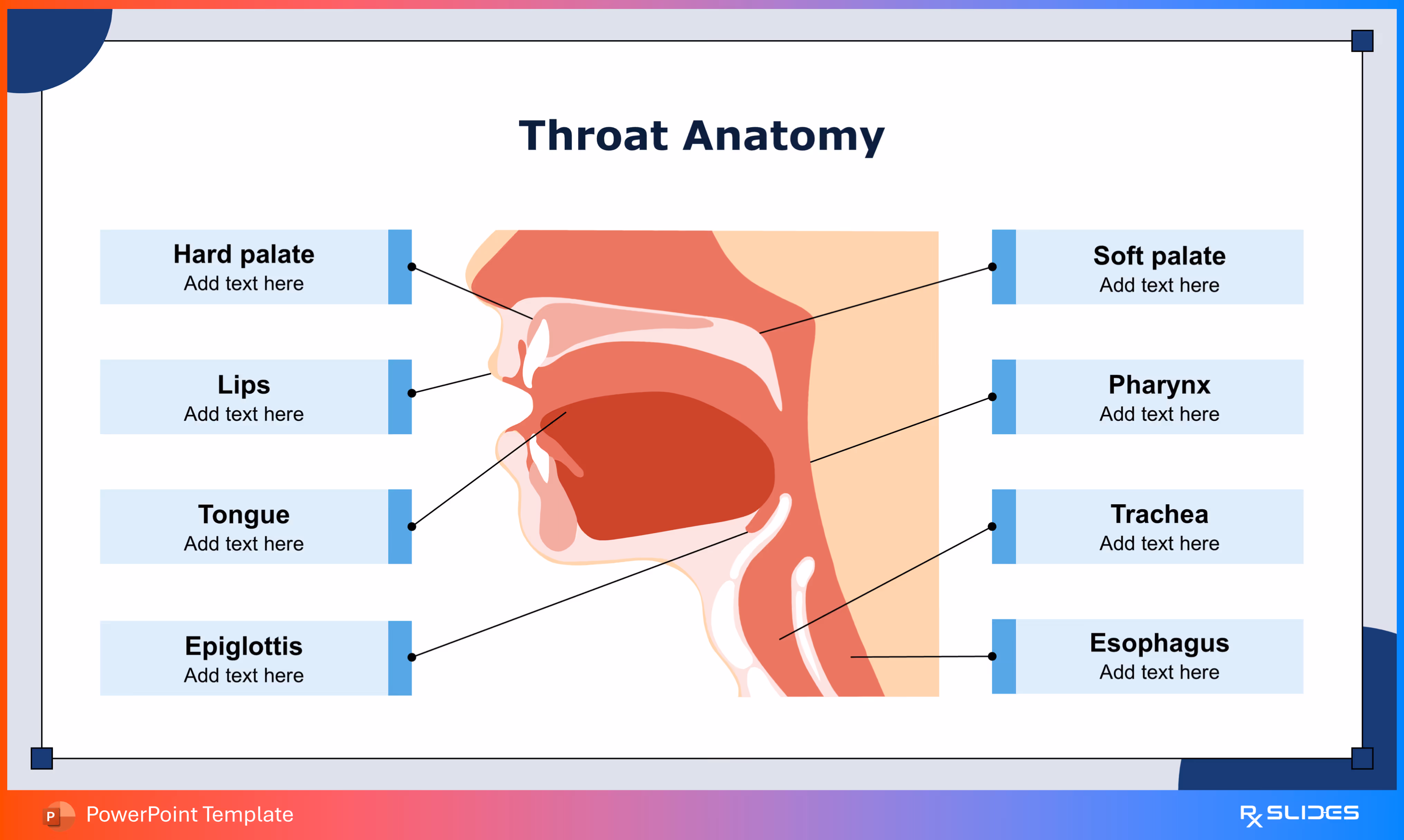
- illustrates the Throat Anatomy relevant to the Epiglottitis presentation.
- The slide features a large, detailed cross-section of the head and throat with eight distinct anatomical features clearly pointed out, including the Epiglottis itself.
Slide 5 - Detailed Throat Anatomy (Epiglottis View)
.avif)
- provides a detailed, close-up look at the Epiglottis and surrounding structures.
- The diagram clearly labels the Epiglottis, Vocal Cords, and the Aryepiglottic Fold.
Slide 6 - Normal Epiglottis Function
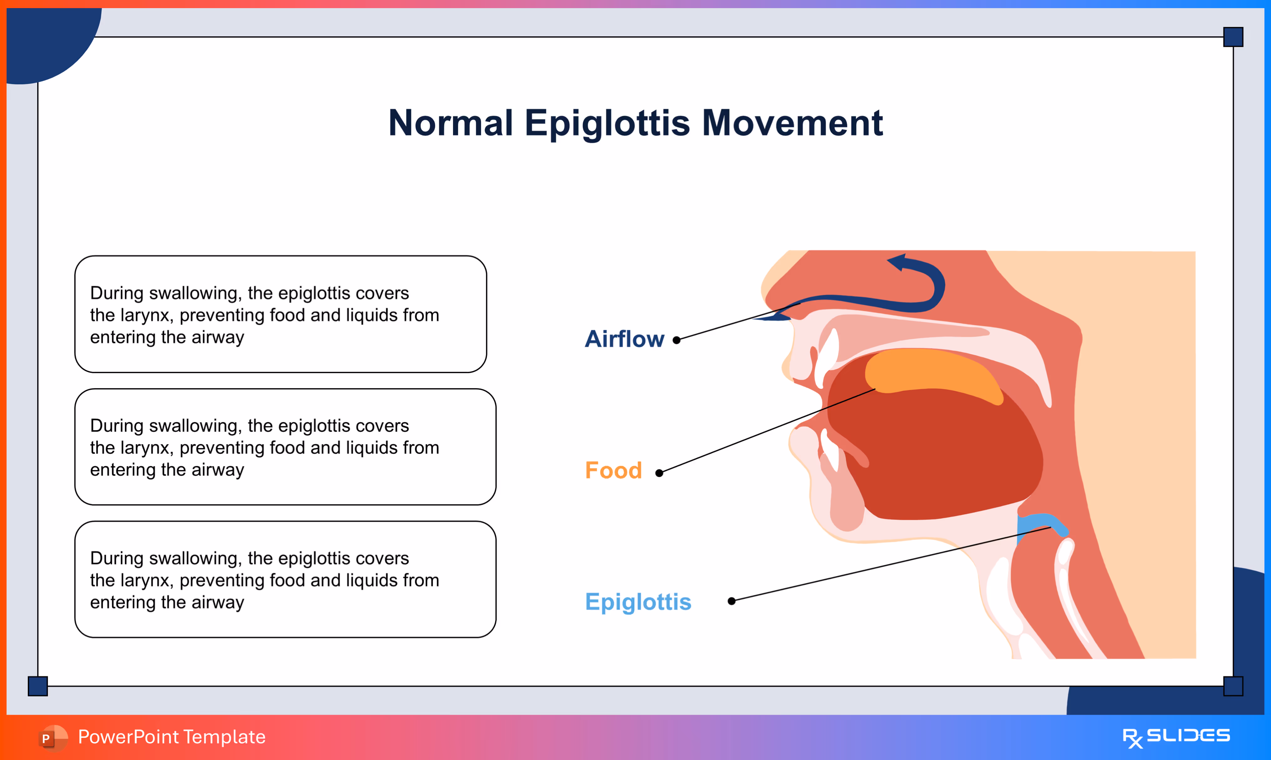
- explains the Normal Movement and crucial protective function of the epiglottis during swallowing.
- The visual highlights the path of Airflow and Food.
Slide 7 - Normal Epiglottis Movement (Breathing)
.avif)
- explains the Normal Movement of the epiglottis when a person is breathing.
- The visual clearly demonstrates the wide open pathway for air into the larynx, contrasting this movement with the protective closure shown in the previous slide.
- This slide clarifies the dual function of the epiglottis.
Slide 8 - Comparison of Normal Epiglottis Movement
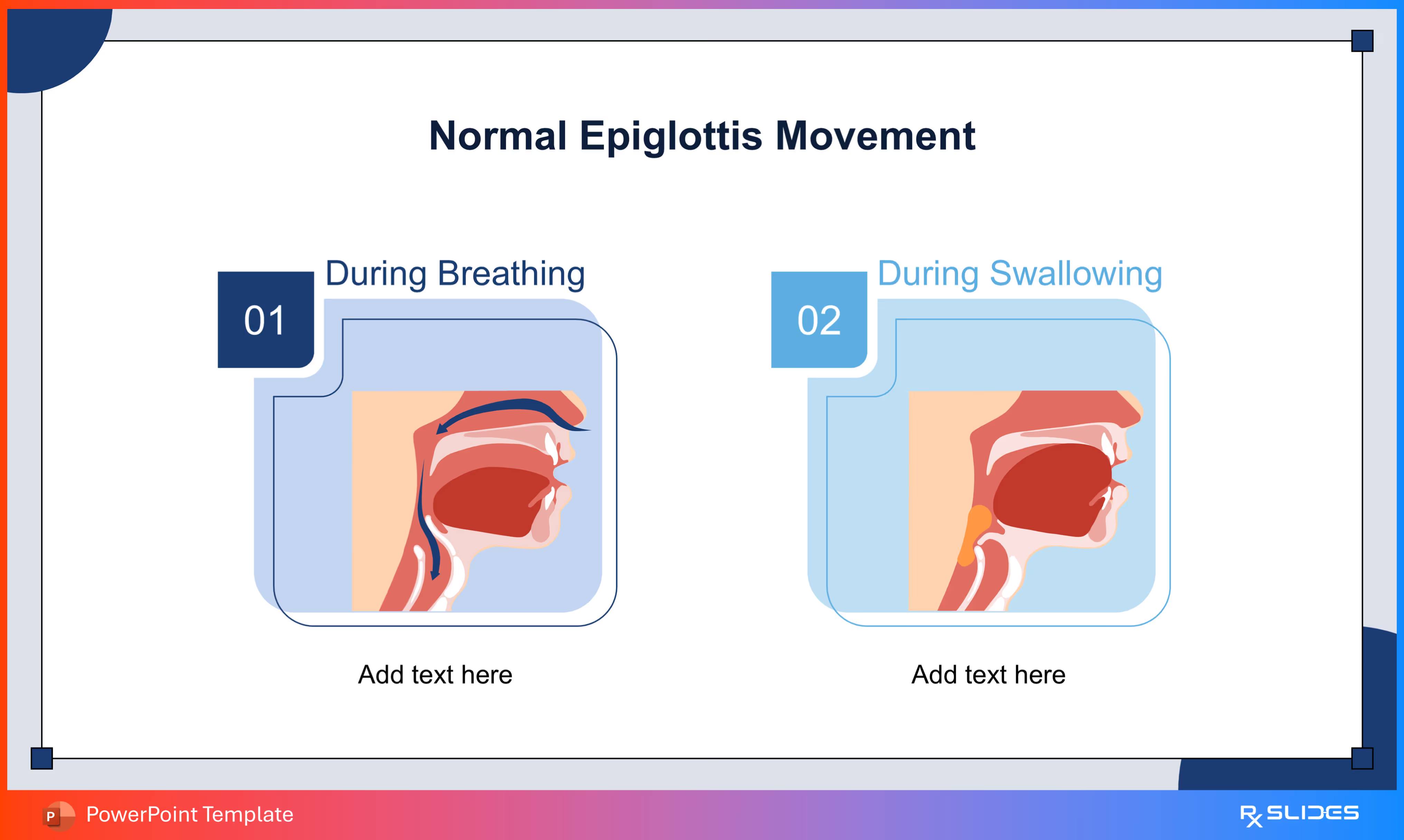
- summarizes the two essential, normal functions of the epiglottis for your audience.
- The visual features two distinct, numbered diagrams (01 and 02) illustrating the open state During Breathing and the closed state During Swallowing.
Slide 9 - Detailed Functional Comparison
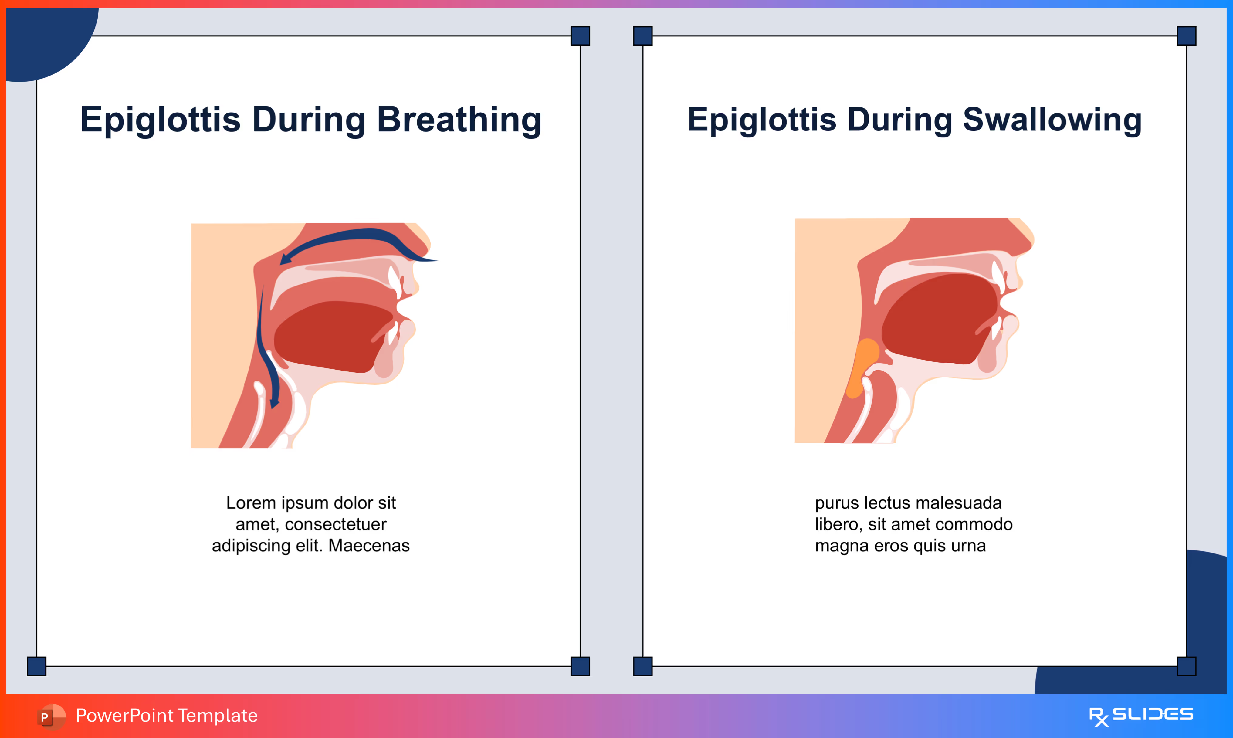
- comparison slide to reinforce the critical difference in epiglottis function between Breathing and Swallowing.
- The slide features large, clear anatomical diagrams placed side-by-side with separate titles.
Slide 10 - Swallowing Phases Breakdown
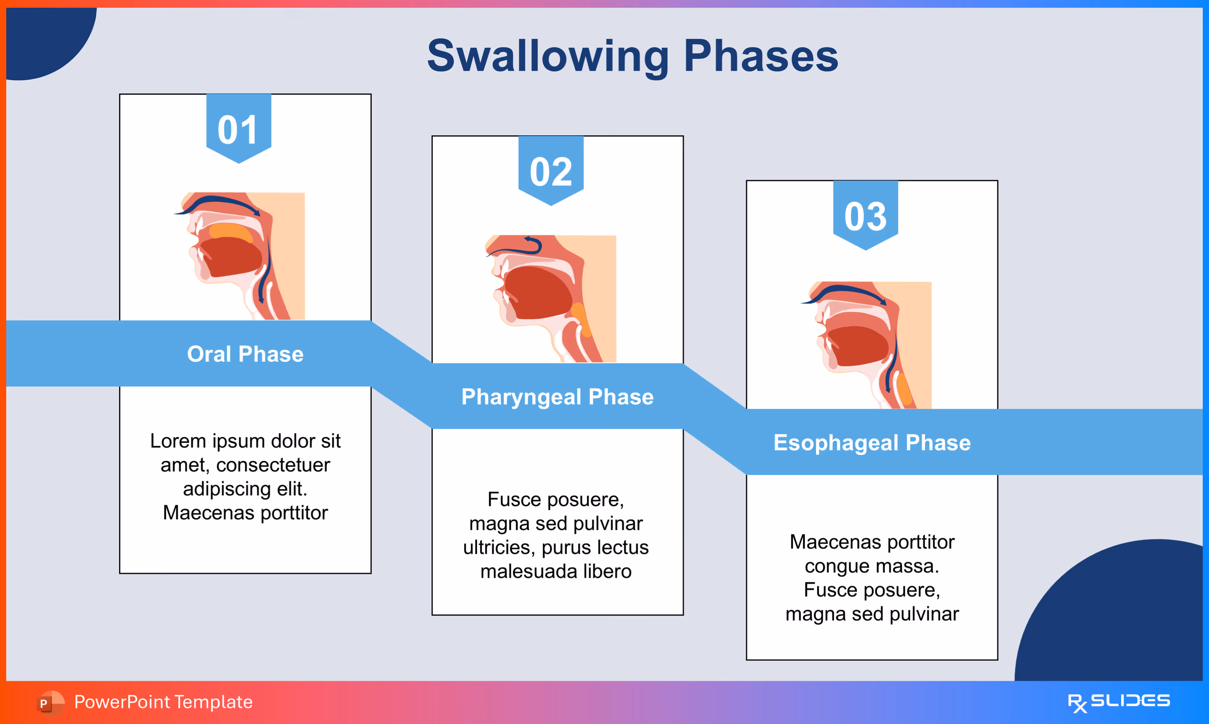
- This slide shows sequential timeline to clearly explain the three distinct Swallowing Phases: Oral, Pharyngeal, and Esophageal.
- The slide features numbered steps (01, 02, 03) corresponding to separate anatomical diagrams.
Slide 11 - Etiology Section Divider
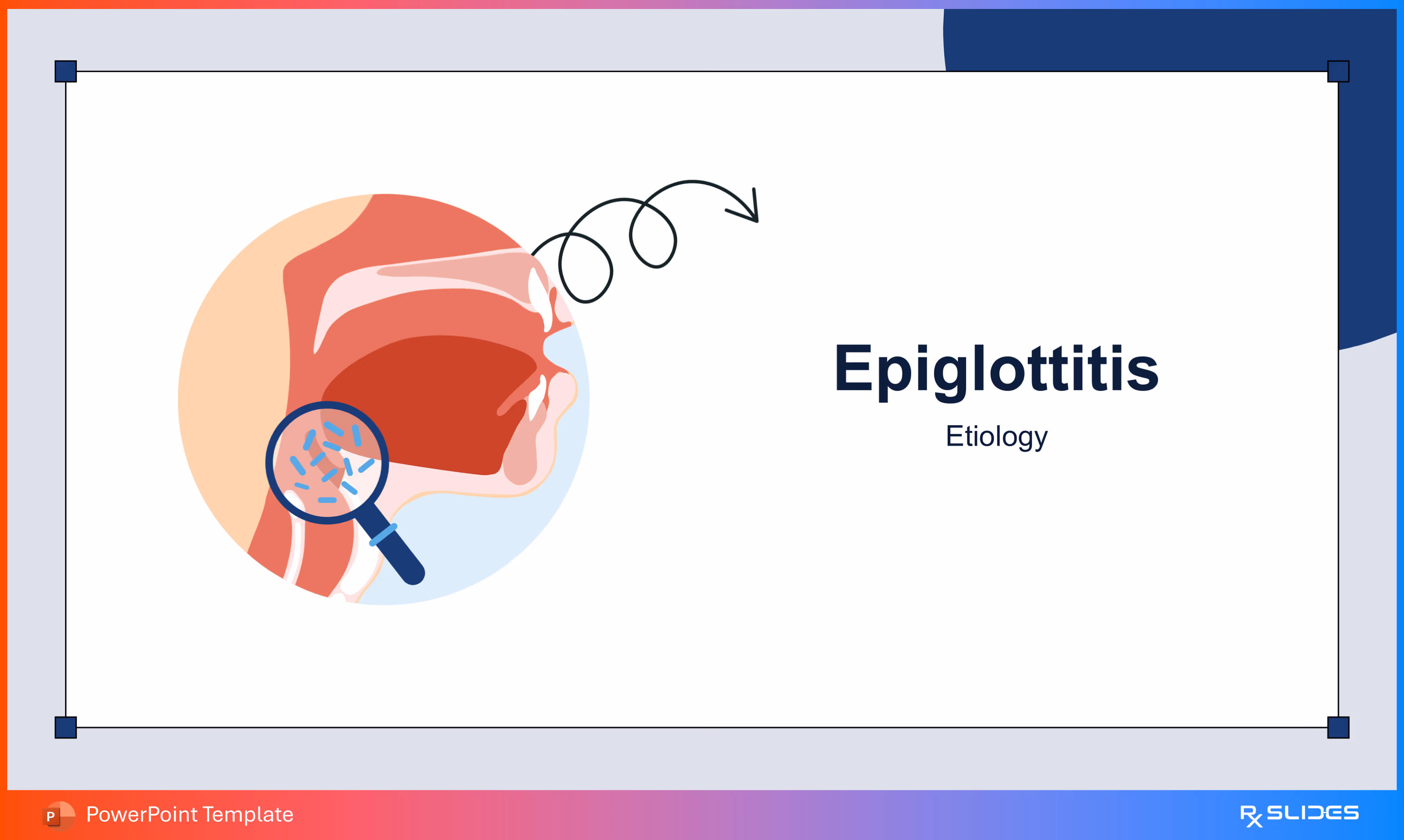
- divider slide to clearly transition to the Etiology (causes) section of your Epiglottitis presentation.
- The visual features a magnified view of the throat, showing bacteria under a magnifying glass, which instantly communicates the infectious origin of the condition.
Slide 12 - Key Etiological Agents
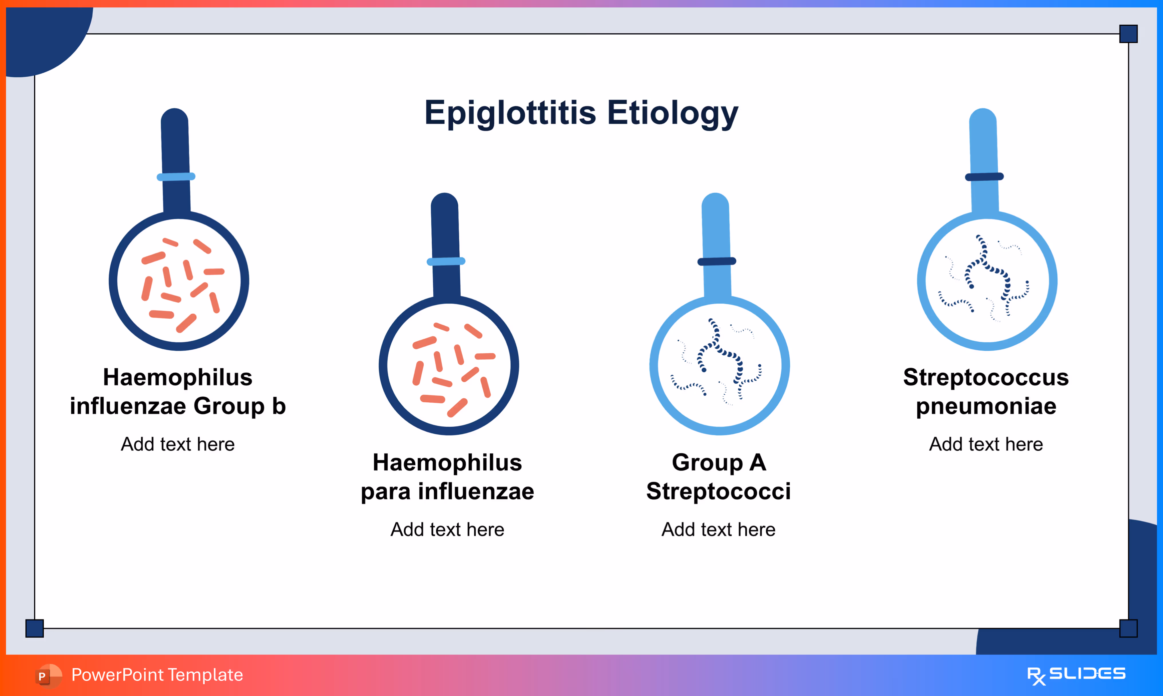
- presents the primary bacteria responsible for Epiglottitis Etiology.
- The slide features four separate magnifying glass icons, each illustrating and labeling a key pathogen, including Haemophilus influenzae and Streptococcus pneumoniae.
Slide 13 - Etiological Agents (Block Style)
.avif)
- presents the four primary bacteria responsible for Epiglottitis Etiology in a highly modern and organized format.
- The slide features four distinct, curved boxes, each showcasing the bacterial illustration and the pathogen's name, such as Haemophilus influenzae and Streptococcus pneumoniae.
Slide 14 - Predisposing Factors Section Divider
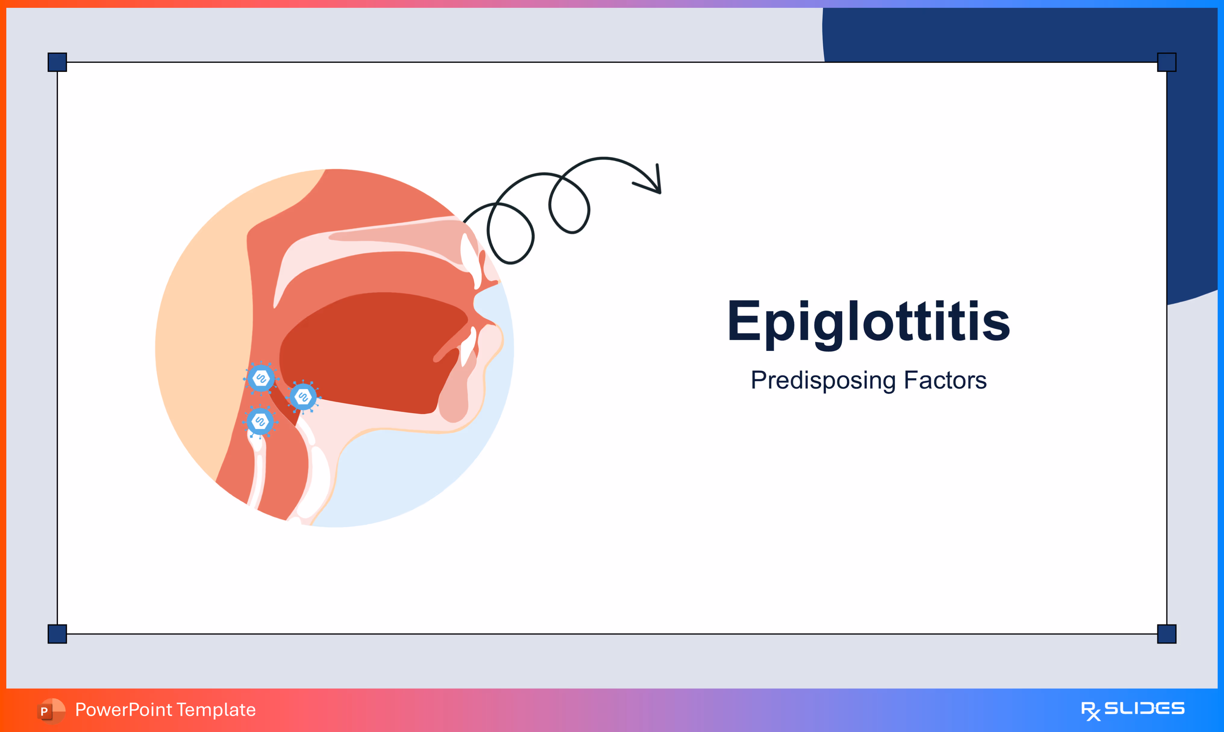
- divider slide to smoothly transition to the section covering the Predisposing Factors of Epiglottitis.
- The visual highlights the throat area with stylized viral icons attached to the epiglottis, suggesting the role of prior conditions or susceptibility in the development of the illness.
Slide 15 - Key Predisposing Factors
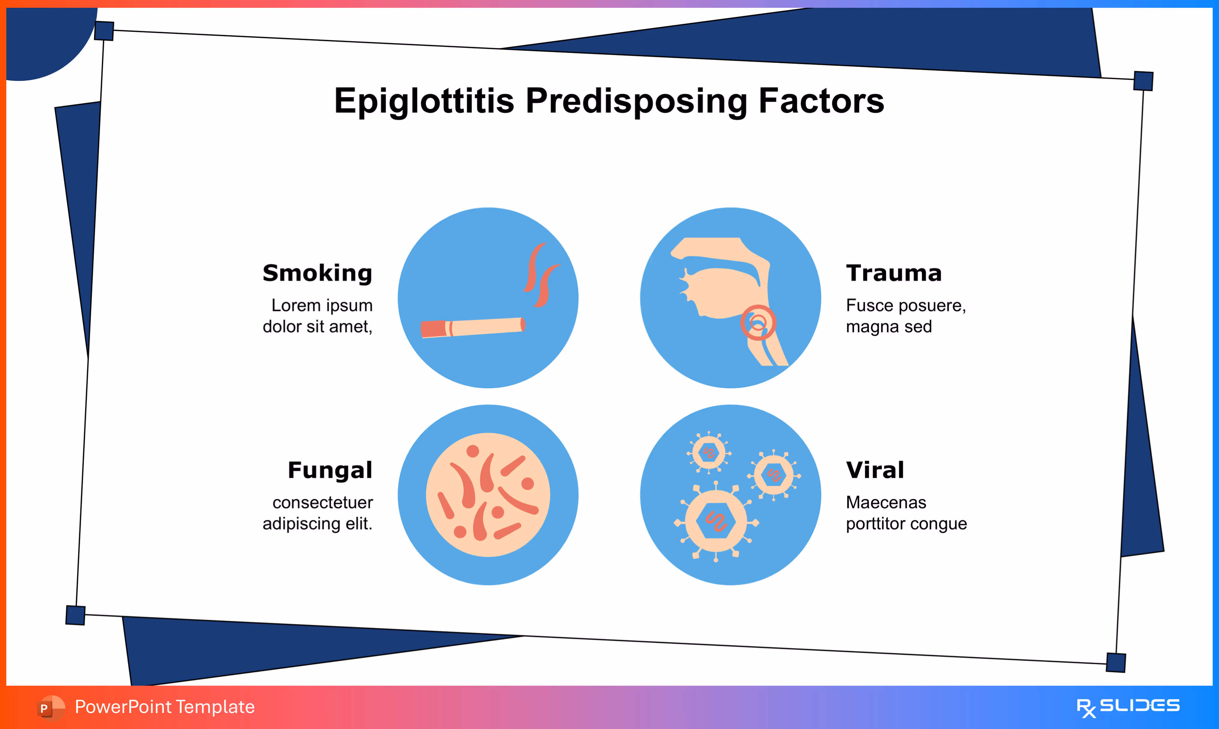
- presents the major Predisposing Factors that increase the risk of developing Epiglottitis.
- The visual includes distinct, easily recognizable icons for factors like Smoking and Trauma, alongside illustrations of Fungal and Viral causes.
Slide 16 - Predisposing Factors (Wheel Layout)
.avif)
- showcases the four primary Predisposing Factors of Epiglottitis in a highly organized and interconnected way.
- The slide divides the information into four clear quadrants, featuring icons for Smoking, Trauma, Fungal infections, and Viral triggers.
Slide 17 - Predisposing Factors (Vertical Timeline)
.avif)
- Presents the four key Predisposing Factors of Epiglottitis in a progressive and structured list.
- The design organizes factors like Trauma and Smoking using distinct icons within circular nodes
Slide 18 - Epidemiology Section Divider
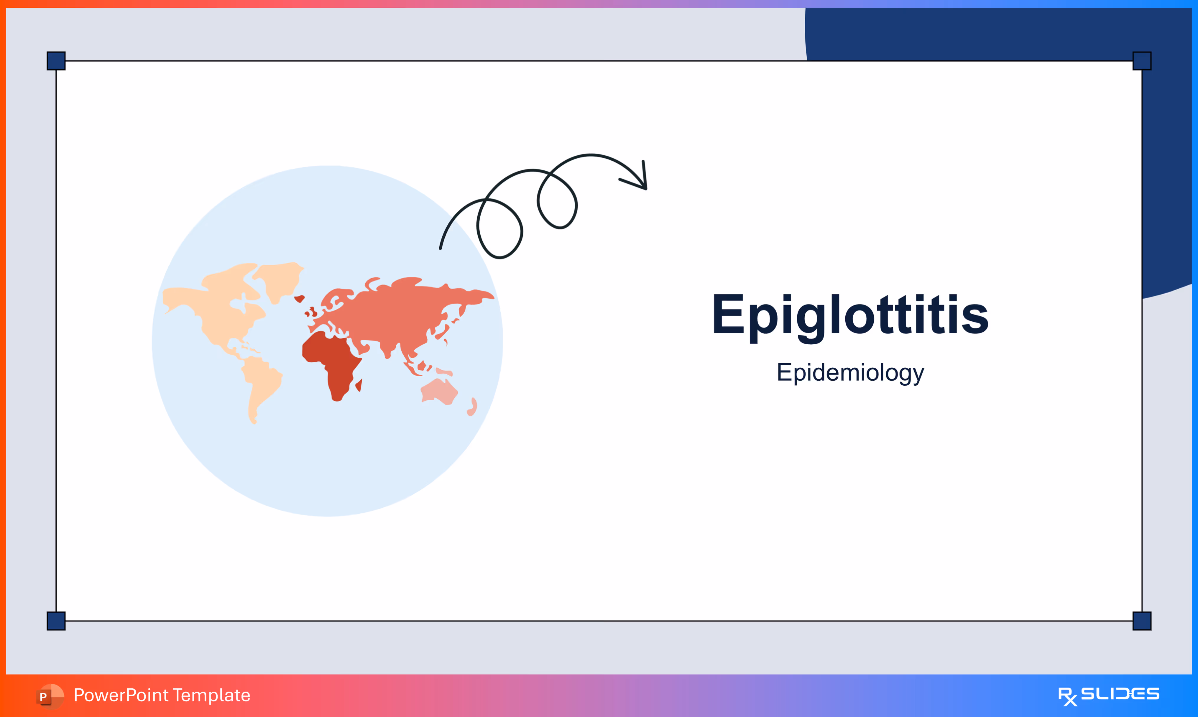
- divider slide to effectively introduce the Epidemiology section, covering the global distribution and study of Epiglottitis.
- The visual prominently features a world map with areas highlighted in varying shades of red, clearly communicating data related to prevalence or incidence.
Slide 19 - Epiglottitis Prevalence Data
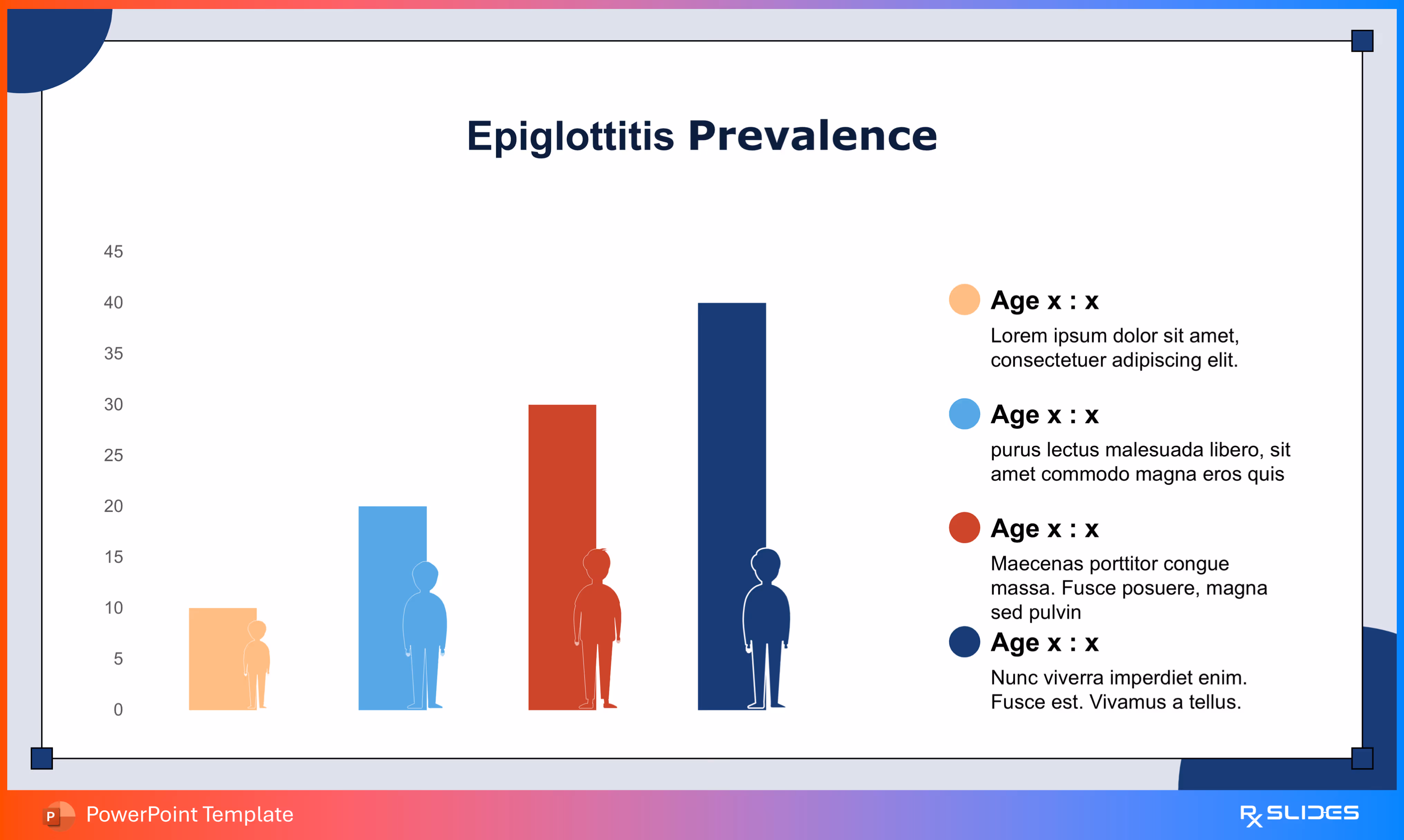
- Shows a four-column bar chart to powerfully present Epiglottitis Prevalence data across different demographic groups or categories.
- The chart utilizes distinct colors and human figures to represent and compare four key data points.
Slide 20 - Epiglottitis Prevalence (Pie Chart)
.avif)
- breaks down Epiglottitis Prevalence data into four distinct categories, likely representing different age groups.
- The chart clearly segments the data using four colors and corresponding human silhouettes.
Slide 21 - Epiglottitis Transmission
- illustrates the two main methods of Epiglottitis Transmission to your audience.
- The visual distinctly separates the concepts of Respiratory droplets (shown via an infected silhouette) and Direct contact (shown via a hand icon).
Slide 22 - Transmission Methods (Dual Boxes)
- Emphasizes the two major ways Epiglottitis Transmission occurs: Respiratory droplets and Direct contact.
- The slide features two distinct, rounded boxes, each containing a dedicated icon (silhouette for breathing, hand for contact).
Slide 23 - Epiglottitis Susceptibility (Age Range)
.avif)
- highlights the primary age group most susceptible to Epiglottitis, spanning from 2 to 7 years.
- The slide features charming child illustrations and large, distinct labels for "2 years" and "7 years," immediately communicating the highest-risk demographic.
Slide 24 - Susceptibility Age Range (Circular Format)
.avif)
- emphasizes the primary age range of Epiglottitis Susceptibility, from two years to seven years.
- The slide uses engaging, separate circular icons with child figures for the "2 years" and "7 years" labels, connected by a bold directional arrow.
Slide 25 - Pathogenesis Section Divider
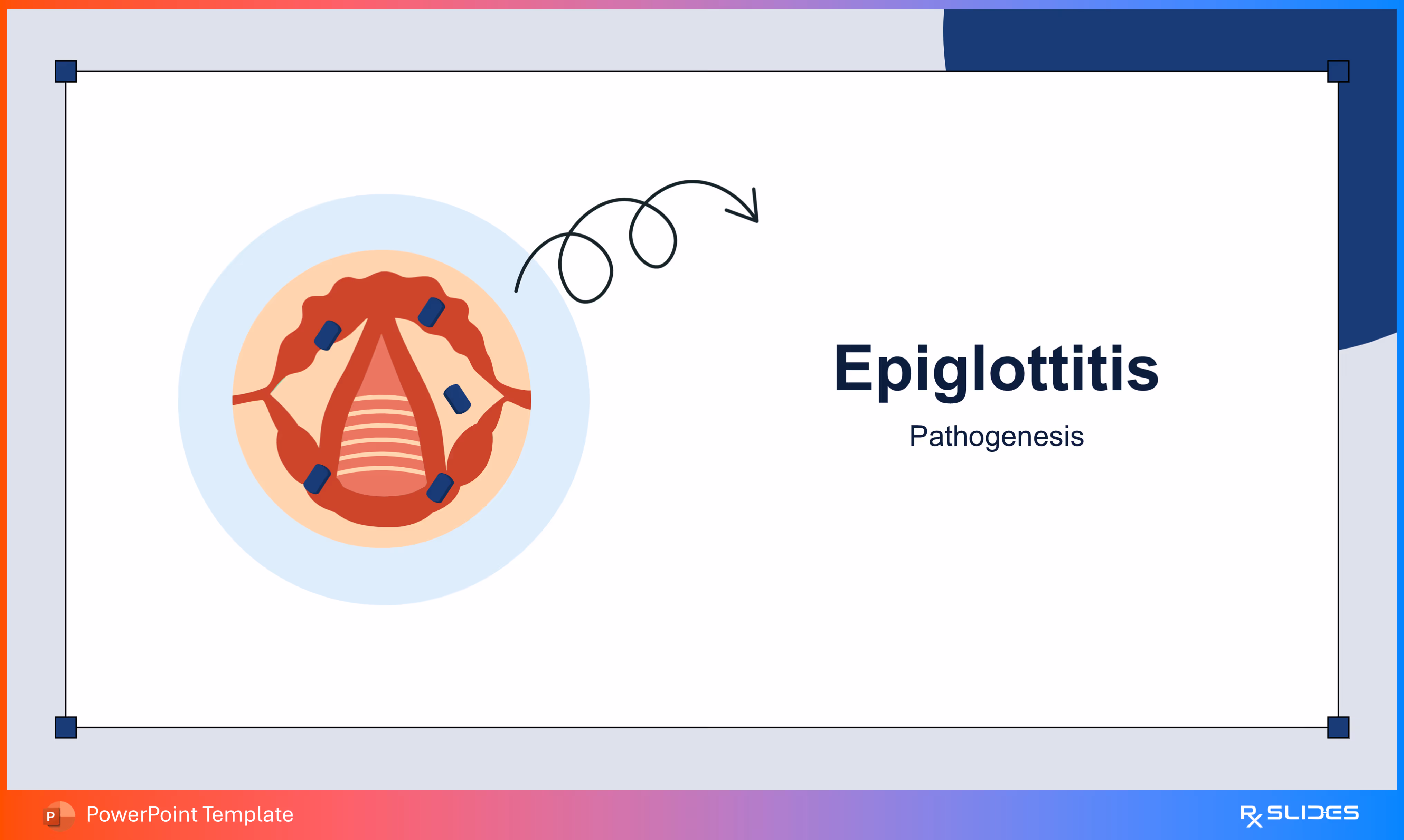
- divider slide to smoothly transition to the Pathogenesis section.
- The visual features a detailed, inflamed view of the epiglottis area, with clear signs of swelling and damage (represented by the dark blue shapes).
Slide 26 - Pathogenesis of Bacterial Epiglottitis
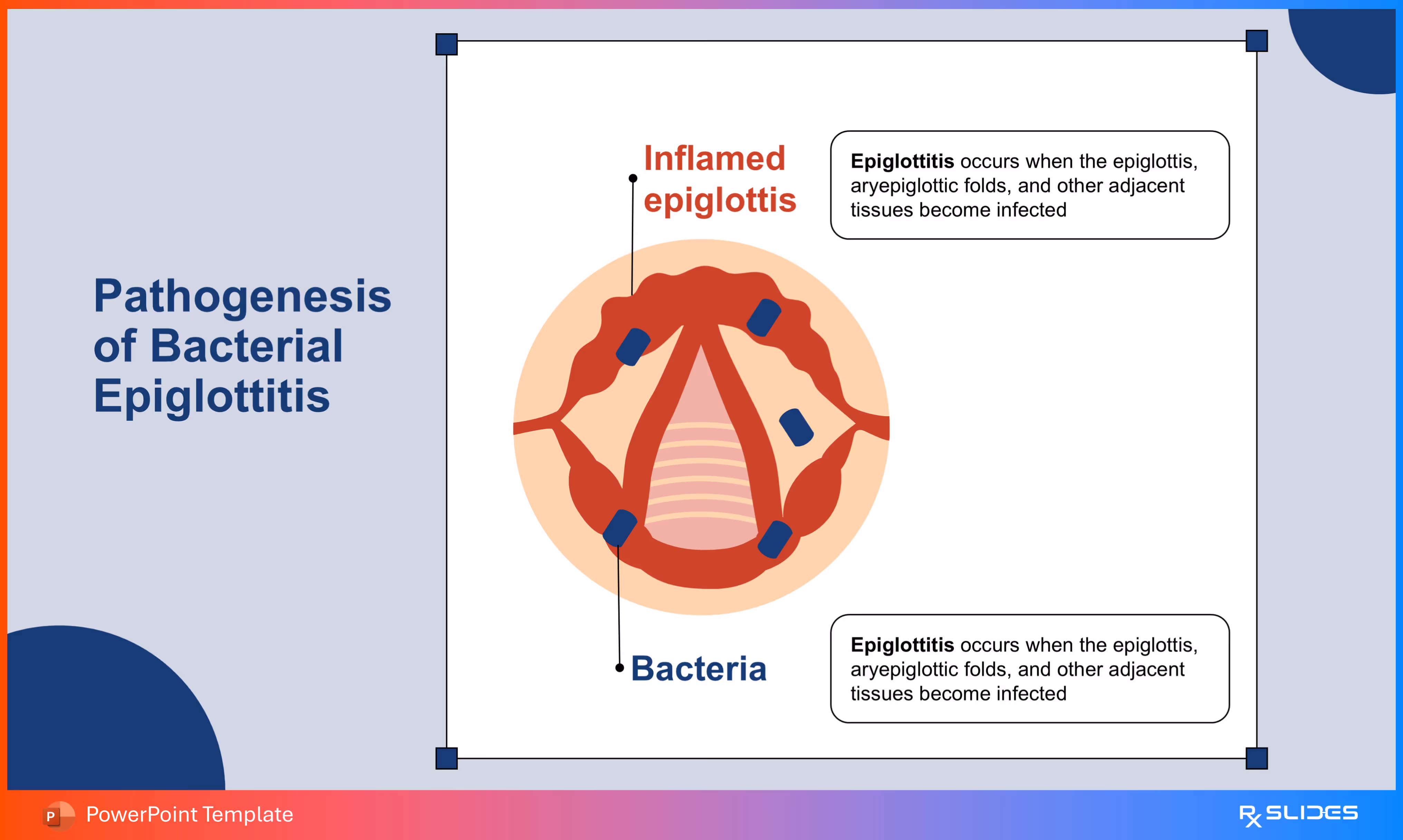
- explains the core mechanism and pathology of Bacterial Epiglottitis.
- The visual highlights the Inflamed epiglottis and surrounding tissues, showing the dramatic swelling and infection caused by the Bacteria (represented by the dark blue shapes).
Slide 27 - Pathogenesis (Cellular Level)
.avif)
- illustrates the initial steps in the Pathogenesis of Bacterial Epiglottitis.
- The visual shows the Bacteria penetrating the Epiglottis Epithelium layer.
Slide 28 - Pathogenesis (Immune Response)
.avif)
- explains the body's Immune Response during the Pathogenesis of Bacterial Epiglottitis.
- The visual highlights how immune cells, such as T-Cells, and inflammatory molecules like TNF-alpha react to the invading bacteria within the tissue layers.
Slide 29 - Pathogenesis (Inflammation Result)
.avif)
- illustrates how inflammation and swelling, the key problems in Epiglottitis, develop at the blood vessel level.
- The visual focuses on a Blood vessel in the epiglottis, demonstrating how inflammatory substances like TNF-alpha increase permeability.
Slide 30 - Pathogenesis (Airway Obstruction)
.avif)
- explains the life-threatening consequence of the Pathogenesis of Bacterial Epiglottitis: airway obstruction.
- The visual features a side-view of the throat showing restricted Airflow due to the Inflamed epiglottis, alongside a diagram showing the dangerous buildup of CO2 in the blood vessels.
Slide 31 - Pathogenesis (Consequence Summary)
.avif)
- shows the two most serious consequences of the Pathogenesis of Bacterial Epiglottitis.
- The slide features the key visual elements: the Inflamed epiglottis restricting Airflow and the Blood vessel showing dangerous CO2 retention.
Slide 32 - Normal vs. Epiglottitis Comparison
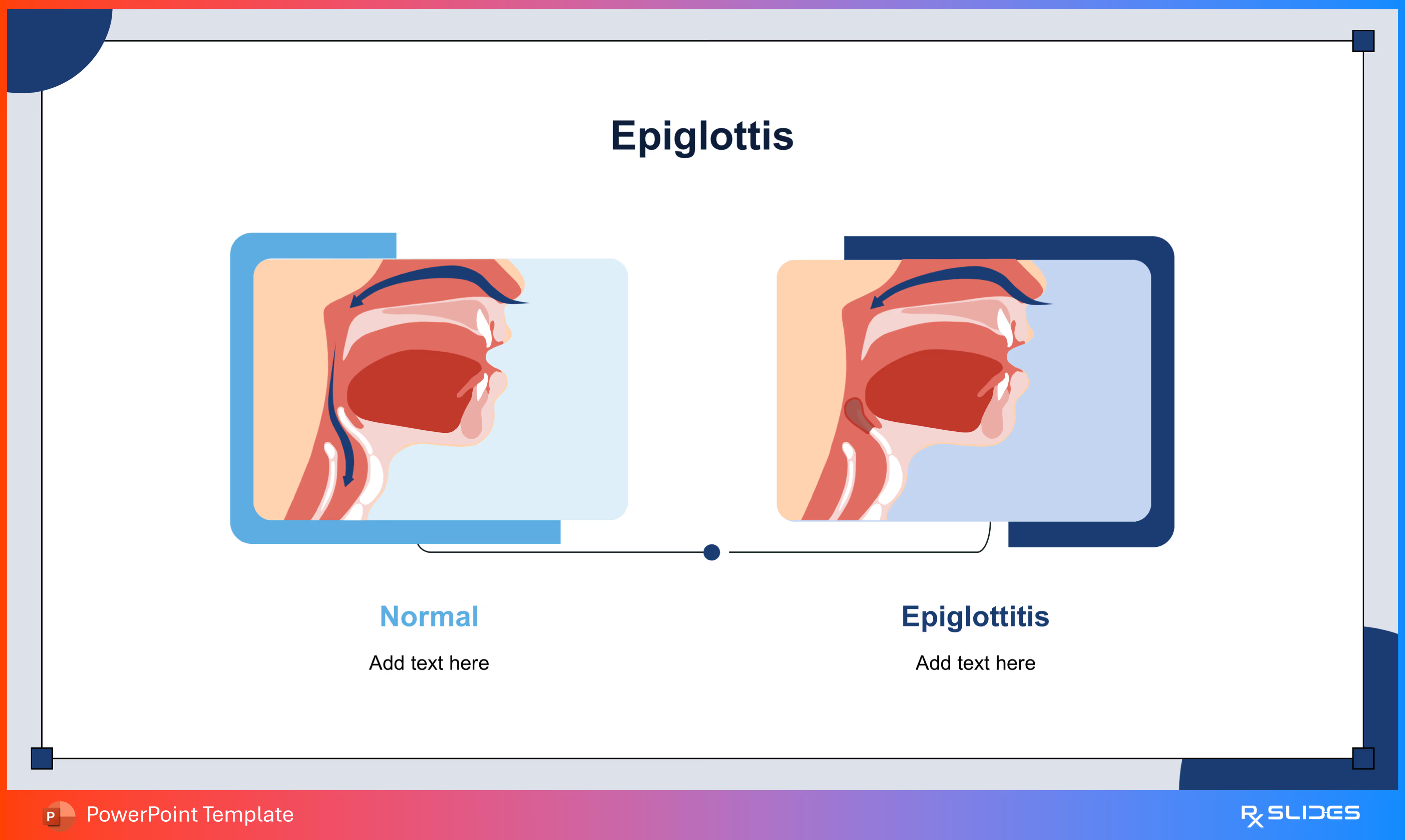
- This slide shows a comparison to instantly demonstrate the difference between the Normal airway and an airway affected by Epiglottitis.
- The slide features two distinct anatomical diagrams contrasting the healthy, open passage on the left with the swollen, obstructed epiglottis on the right.
Slide 33 - Symptoms Section Divider
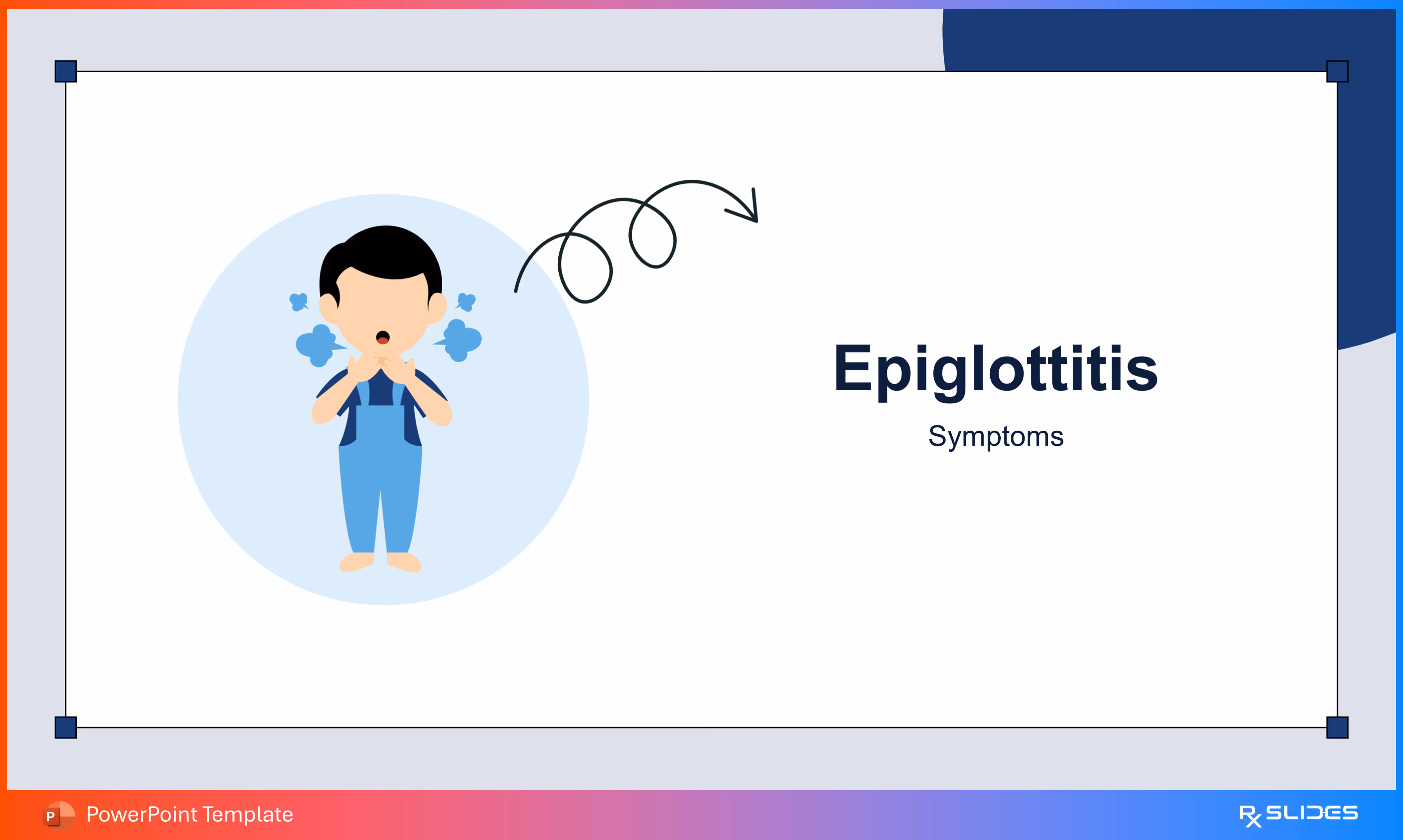
- divider slide to smoothly transition to the section covering the Symptoms of Epiglottitis.
- The visual features an illustration of a child having difficulty breathing (suggesting distress and coughing).
Slide 34 - Key Epiglottitis Symptoms
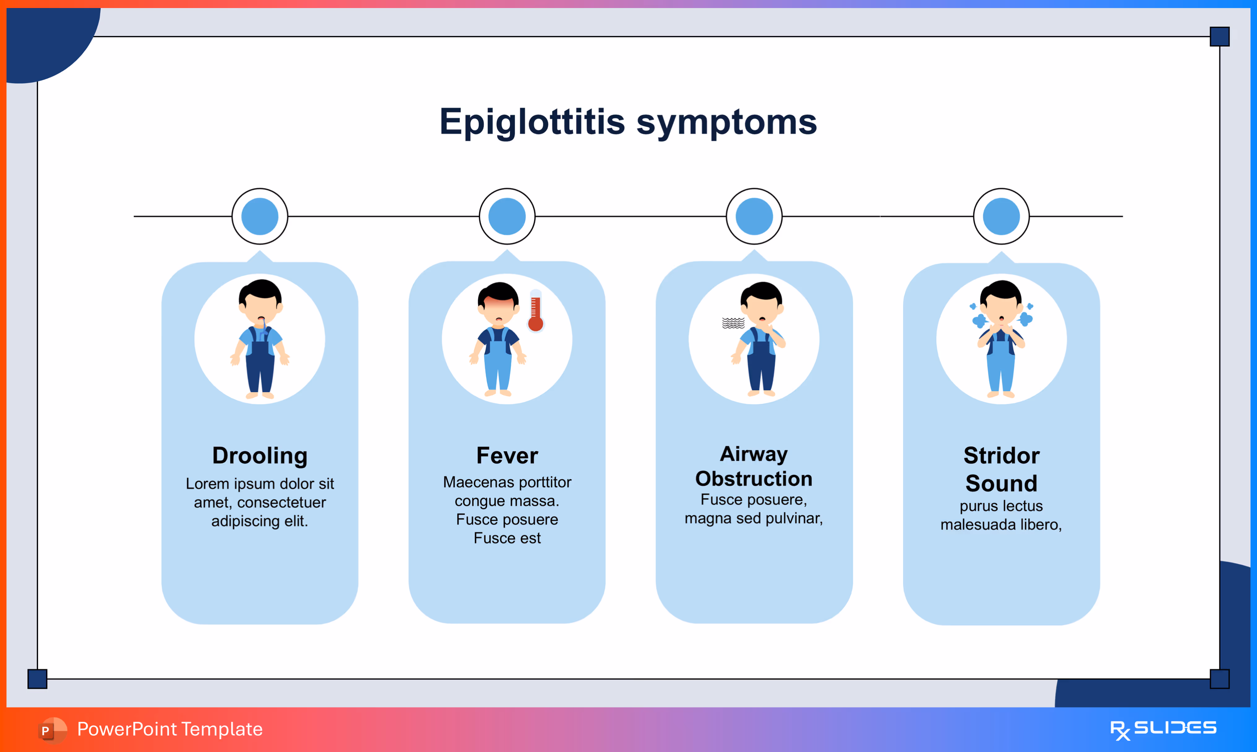
- presents the four most critical Symptoms of Epiglottitis.
- The slide uses dedicated visual icons for each symptom, clearly illustrating key signs like Fever, Drooling, Airway Obstruction, and the Stridor Sound.
Slide 35 - Epiglottitis Symptoms (Boxed Icons)
.avif)
- presents the four key Symptoms of Epiglottitis, giving equal visual weight to each point.
- The slide features four dedicated boxes with clear icon illustrations for Drooling, Fever, Airway Obstruction, and the Stridor Sound.
Slide 36 - Diagnosis Section Divider
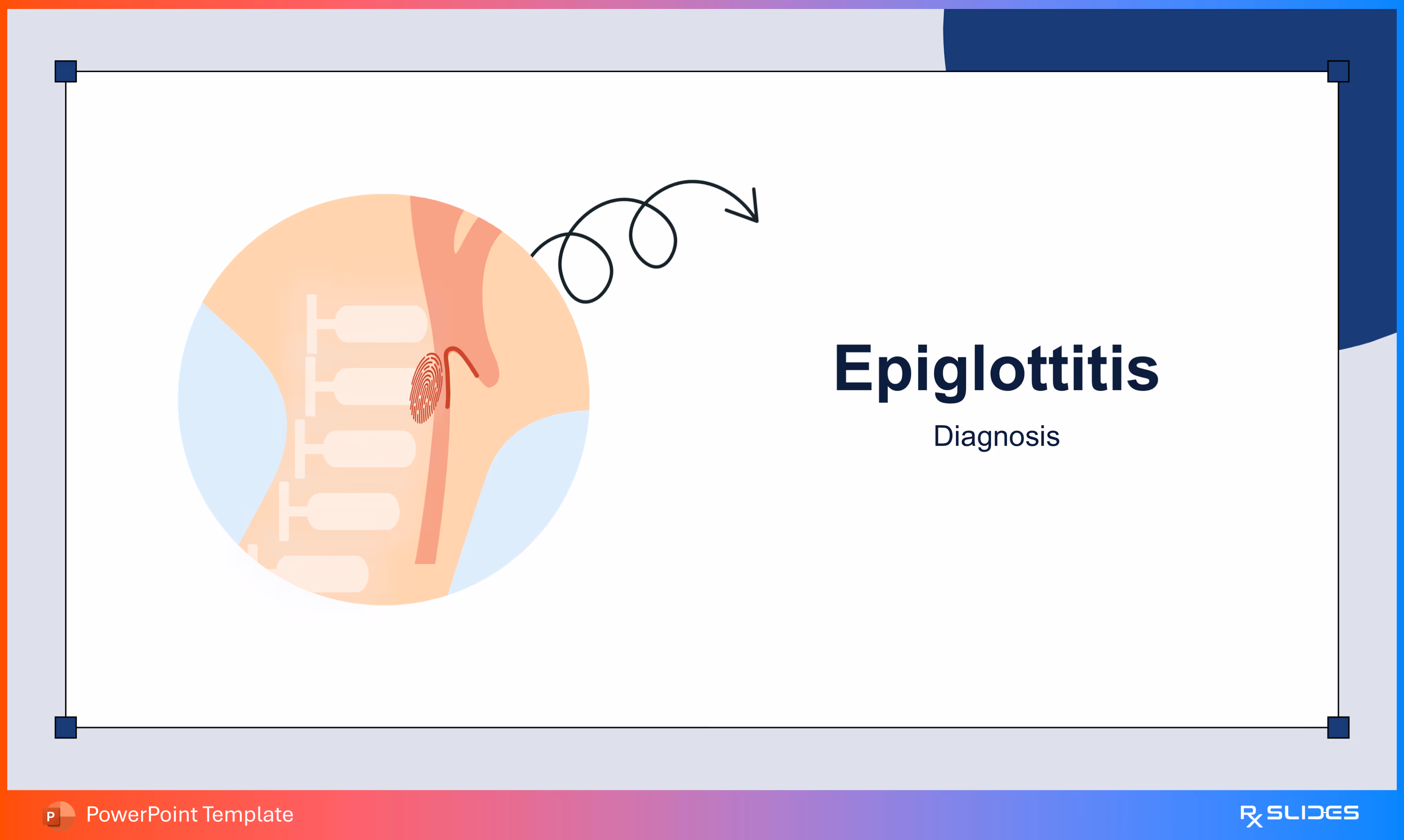
- divider slide to smoothly transition to the Diagnosis section, covering the methods used to identify Epiglottitis.
- The visual features a diagram of the neck and throat area, highlighting the swollen epiglottis adjacent to the vertebrae.
Slide 37 - Diagnosis via X-ray (Thumbprint Sign)
.avif)
- explains how an X-ray confirms Epiglottitis using the distinctive "thumbprint sign."
- The visual features a diagram of the swollen epiglottis next to the vertebrae, with the epiglottis itself stylized like a thumbprint to represent the finding on a lateral X-ray.
Slide 38 - Diagnosis via CT Scan
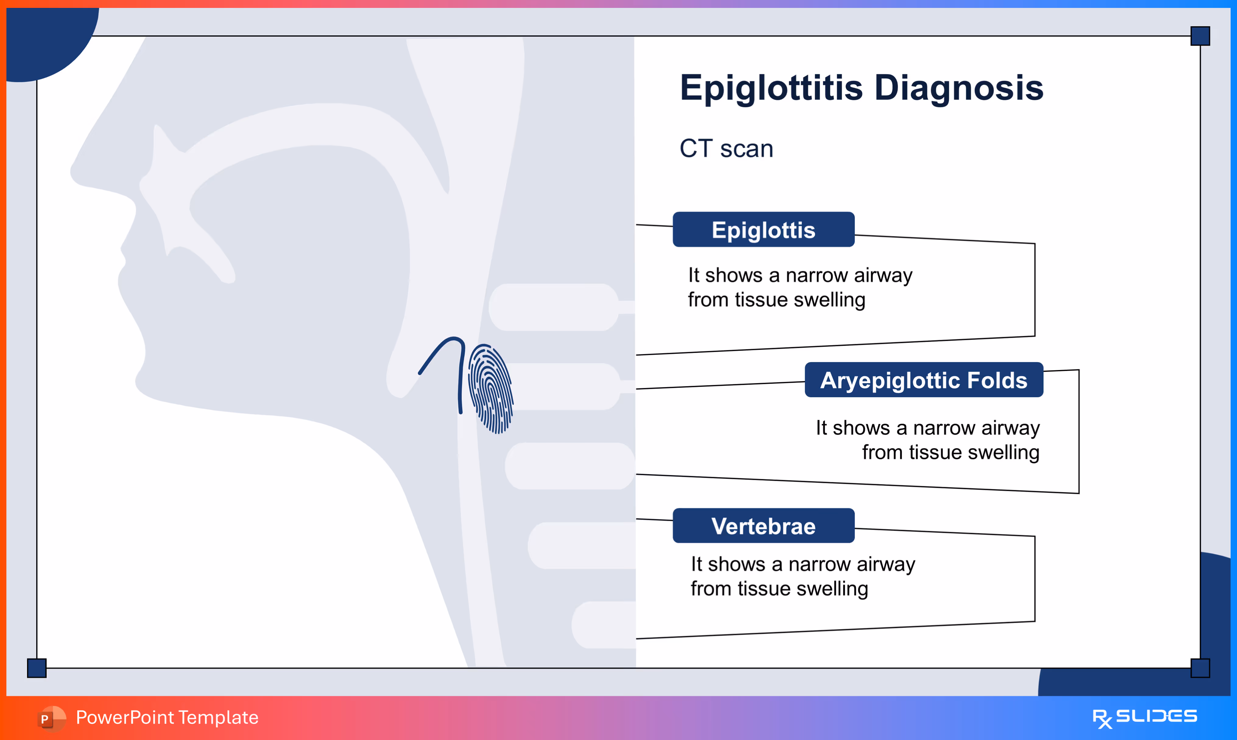
- explains how a CT Scan is used to confirm Epiglottitis by identifying airway narrowing.
- The visual highlights the Epiglottis and surrounding Aryepiglottic Folds.
Slide 39 - Diagnosis via Endoscopy
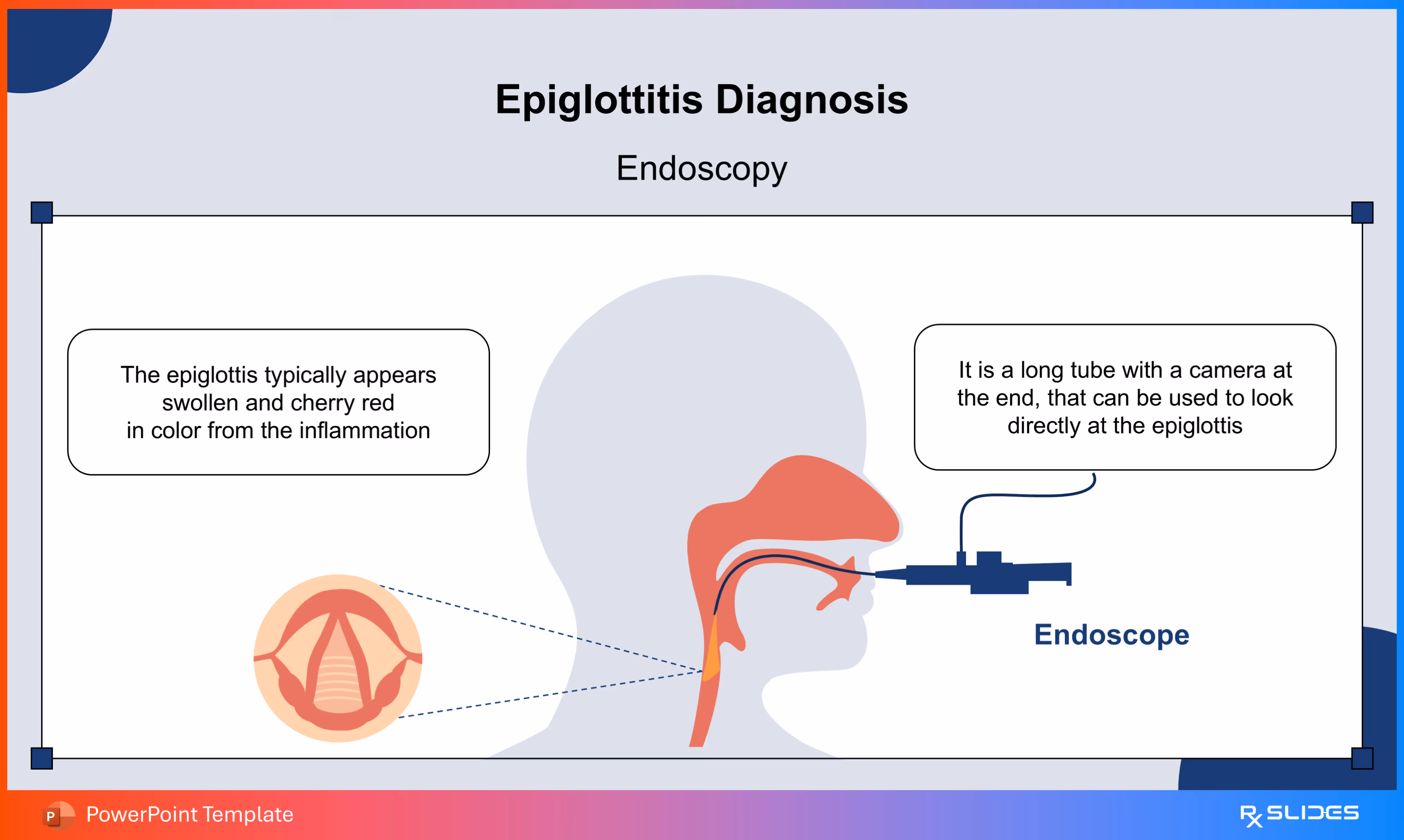
- explains the procedure and findings of Endoscopy, a key technique for diagnosing Epiglottitis.
- The visual shows an Endoscope being inserted into the throat, with a magnified inset illustrating the typical swollen, cherry-red appearance of the inflamed epiglottis.
Slide 40 - Treatment Section Divider
- divider slide to smoothly transition to the Treatment section.
- The visual features a striking illustration of a resuscitation bag or Ambu-bag.
Slide 41 - Treatment Options Overview
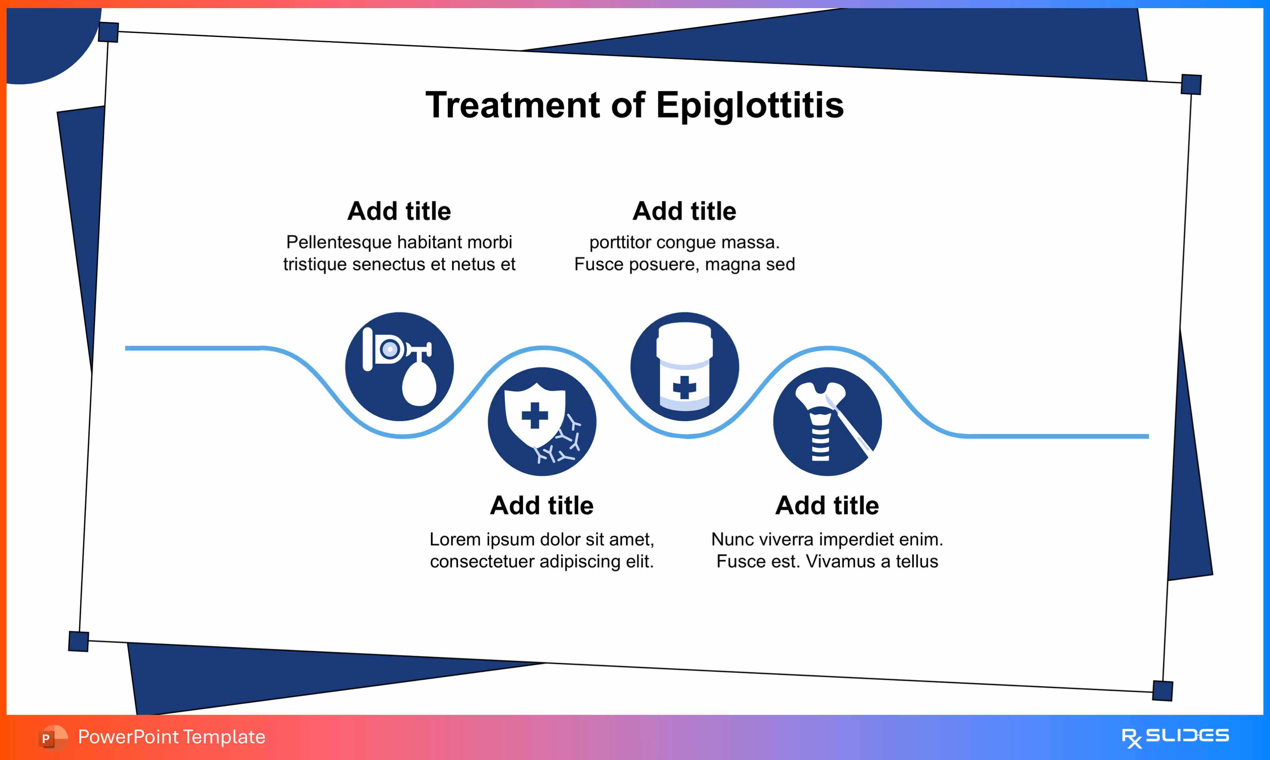
- summarizes the major Treatment Options for managing Epiglottitis.
- The slide features four distinct icons placed along a wave-like line, representing key areas like Airway Management, Antibiotics, and potentially Vaccination or supportive care.
Slide 42 - Key Epiglottitis Treatment Methods
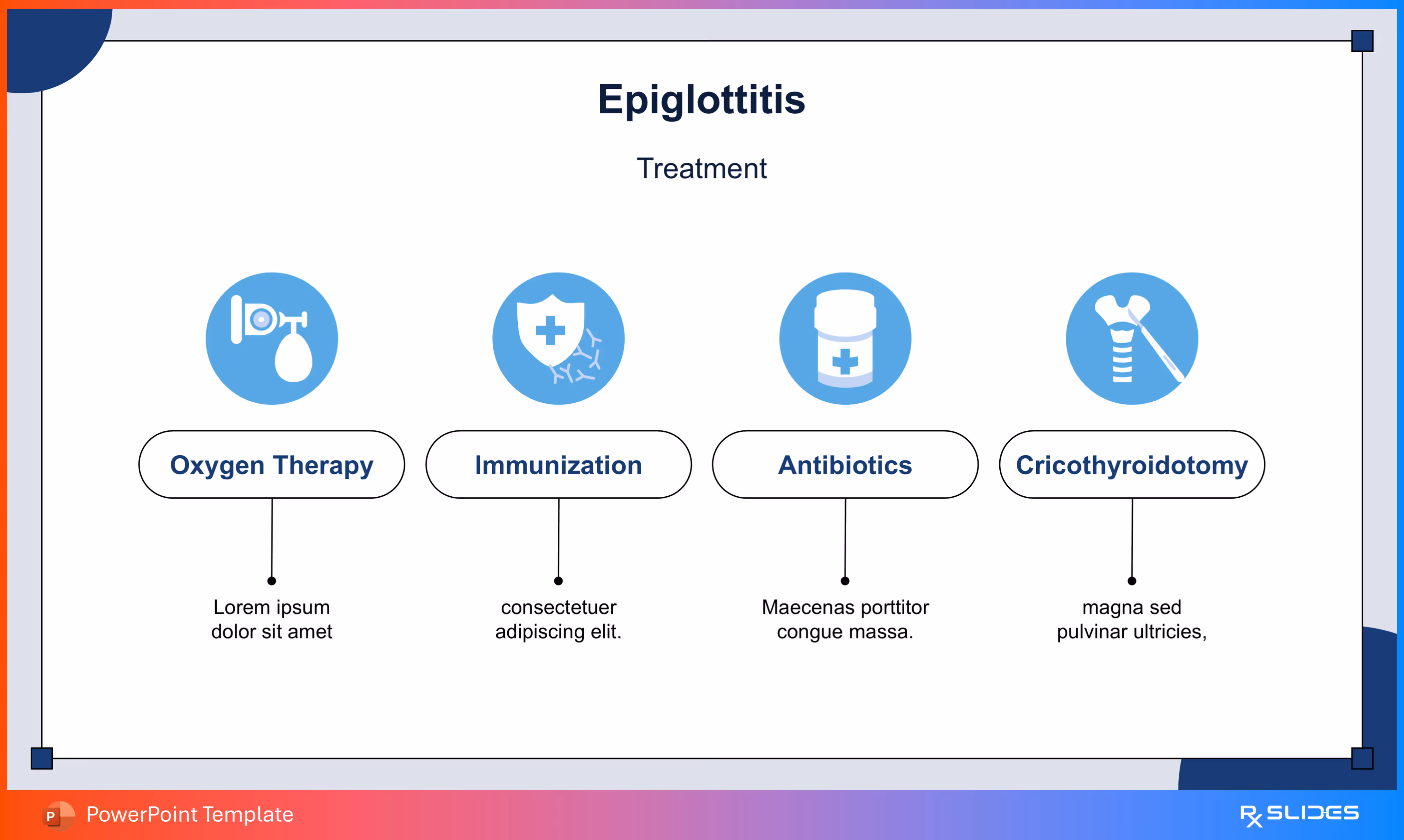
- presents the four essential Treatment Methods for managing Epiglottitis.
- The slide features dedicated icons and clear labels for Oxygen Therapy, Immunization, Antibiotics, and Cricothyroidotomy, covering the critical steps from supportive care to emergency surgery.
Slide 43 - Alternative Treatment Slide
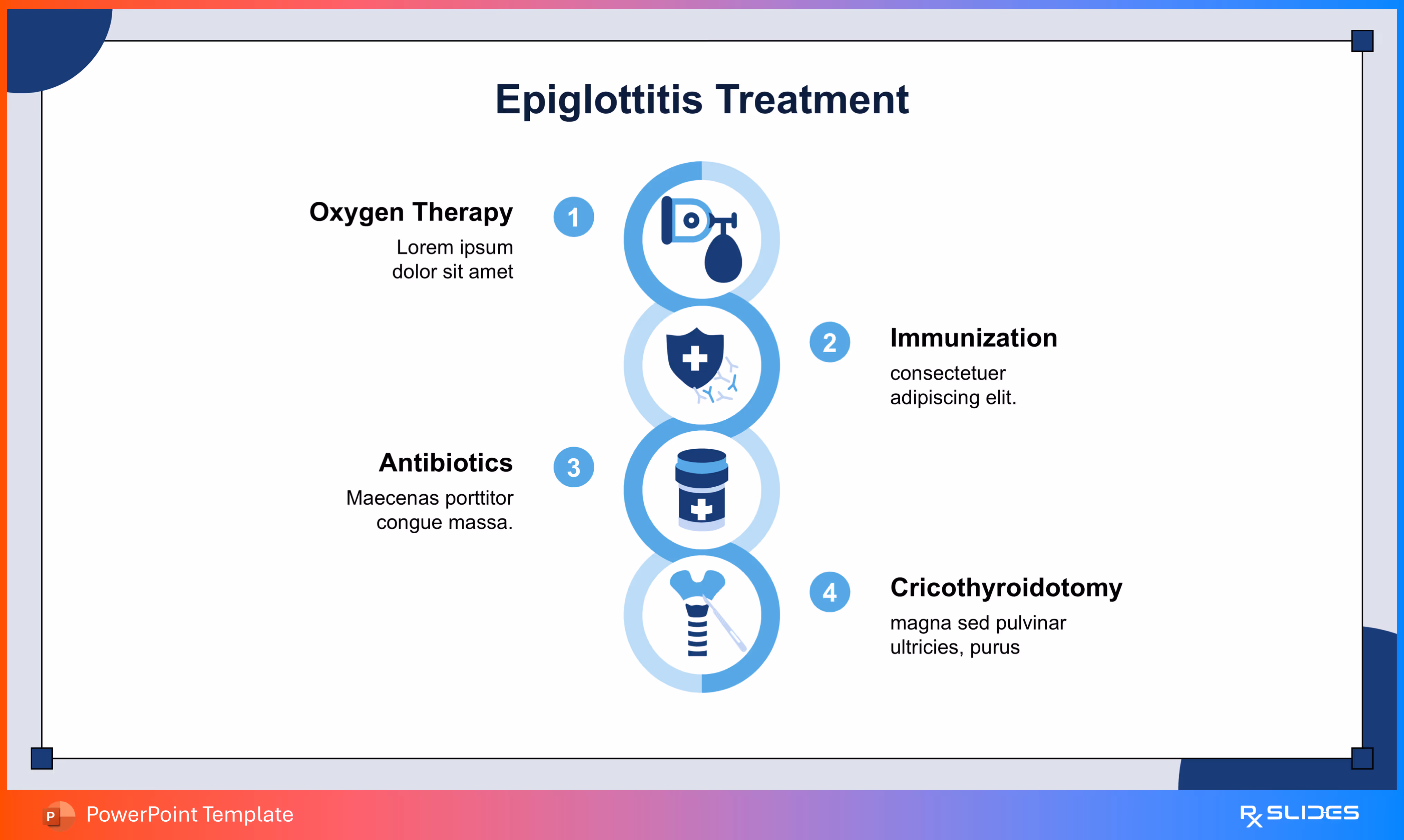
- 43 slide is an alternative slide for 42
Slide 44 - Endotracheal Intubation
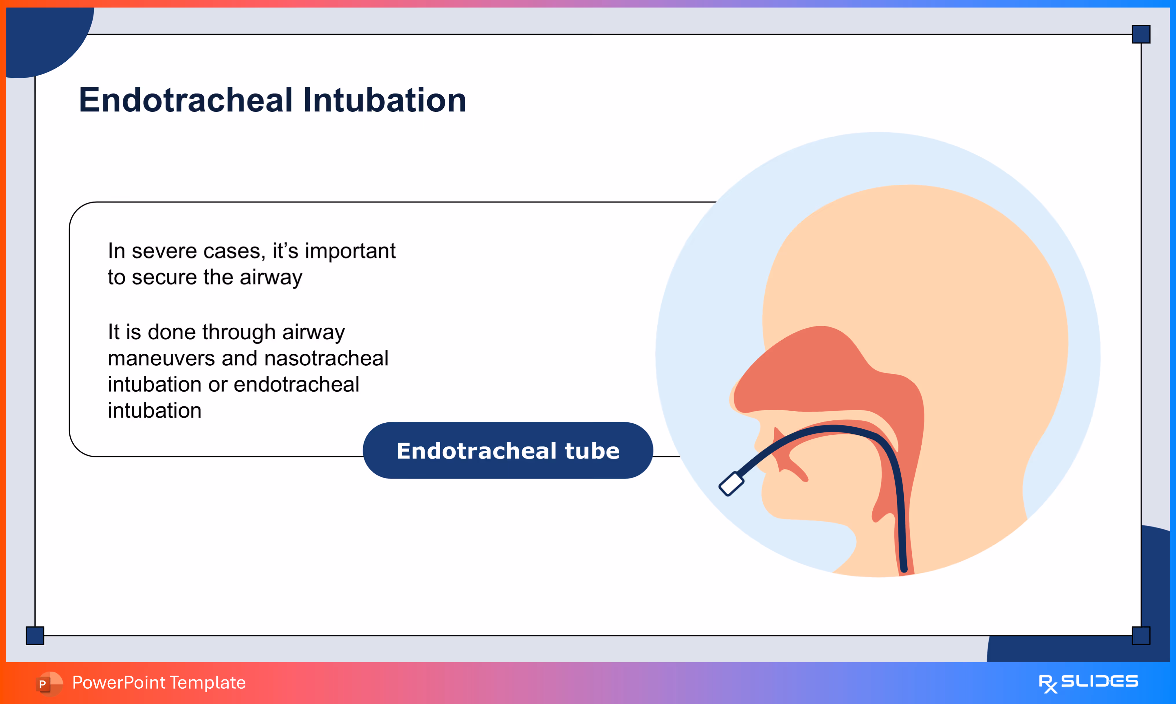
- explains the process of Endotracheal Intubation, the key procedure for securing the airway in severe Epiglottitis.
- The visual features a side-view of the throat showing the Endotracheal tube being inserted past the epiglottis to ensure continuous, safe airflow.
Slide 45 - Prevention Section Divider
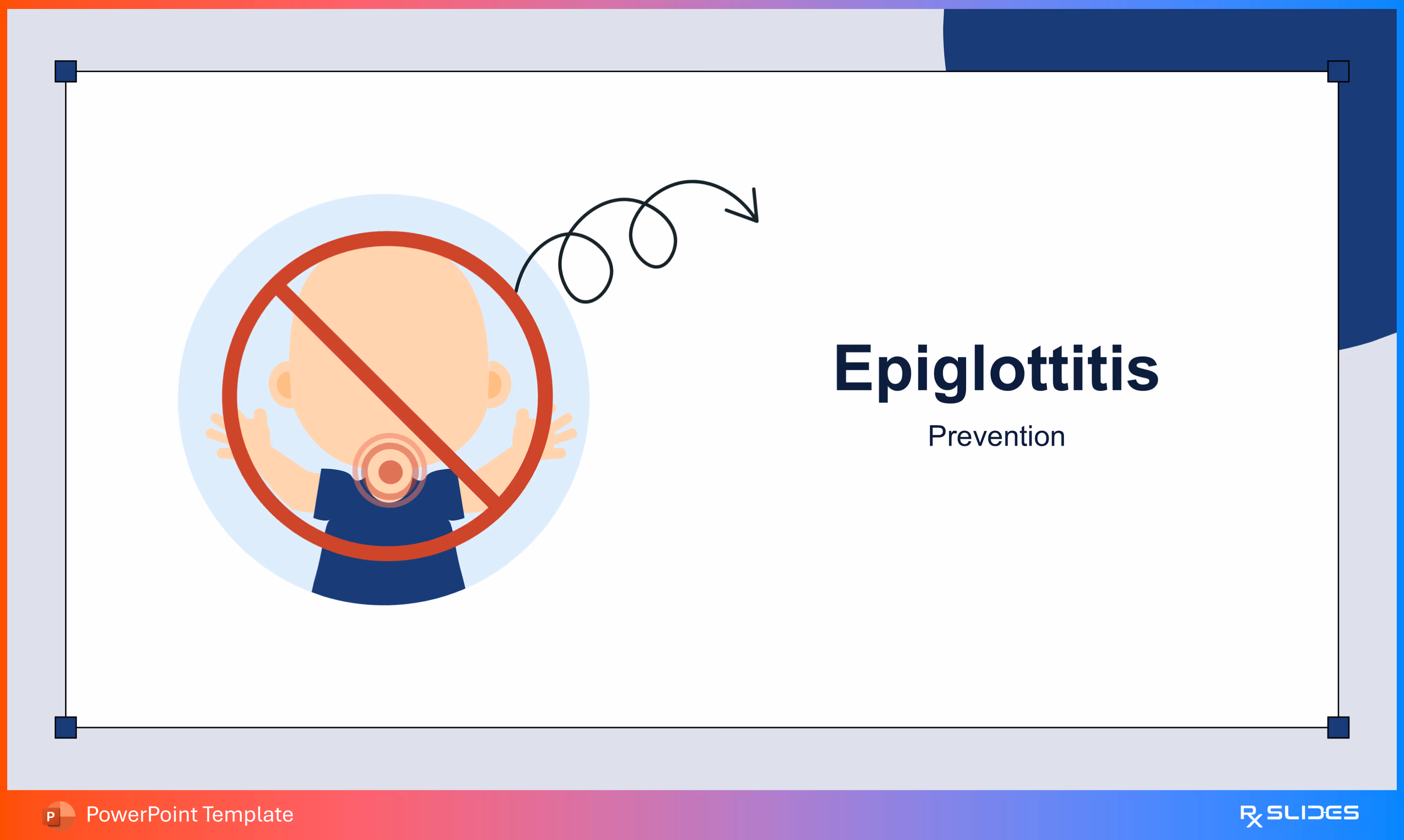
- divider slide to smoothly transition to the Prevention section.
- The visual features a striking illustration of a child with a red circle and line (a "no" symbol) over the throat.
Slide 46 - HiB Vaccination Program (Prevention)
.avif)
- presents the critical HiB Vaccination Program schedule.
- The visual features a cute baby alongside four distinct boxes detailing the recommended administration months: 2 Months, 4 Months, 6 Months, and 12 Months.
Slide 47 - HiB Vaccination Schedule (Circular Layout)
.avif)
- presents the four recommended doses of the HiB Vaccination Program.
- The slide features a prominent illustration of a syringe and vaccine vial next to four large, color-coded circles marking the critical vaccination months: 2, 4, 6, and 12 months.
Slide 48- HiB Vaccination Schedule (Three-Dose Layout)
.avif)
- presents a slightly simplified version of the HiB Vaccination Program, focusing on the 2 month, 4 month, and 12 month doses.
- The slide features three charming baby icons, each sitting above a blue block indicating the crucial administration time points.
Slide 49 - HiB Vaccination Program (Alternate Three-Dose Layout)
.avif)
- highlights the key time points of the HiB Vaccination Program for Epiglottitis prevention.
- The slide features three cute, expressive baby figures placed below three distinct, large blue boxes that specify the crucial administration times: 2 months, 4 months, and 12 months.
Slide 50 - Thank You/Conclusion Slide

- concludes the presentation with a thank you message.
- The visual features a large, clear "Thank You" title alongside the prominent throat anatomy graphic.
Features of the Template
- 100% editable PowerPoint template.
- Editable colors, you can change according to your presentation style and company branding guidelines.
.avif)


.avif)