How to Use Animation to Explain Mechanisms of Action in your Presentation
The most effective way to explain Mechanisms of Action (MoA) in a PowerPoint presentation is through the use of animated medical slides. These dynamic visuals are specifically designed to transform complex medical concepts into clear, engaging, and scientifically accurate narratives, ensuring that the audience understands even the most intricate details of a drug's action or a disease's pathophysiology.
RxSlides templates, designed by medical professionals, utilize specific animation techniques to visualize MoA, progressing from molecular binding to systemic effects.
Here is how animation is used to explain mechanisms of action:
1. Illustrating Cellular and Receptor Cascades
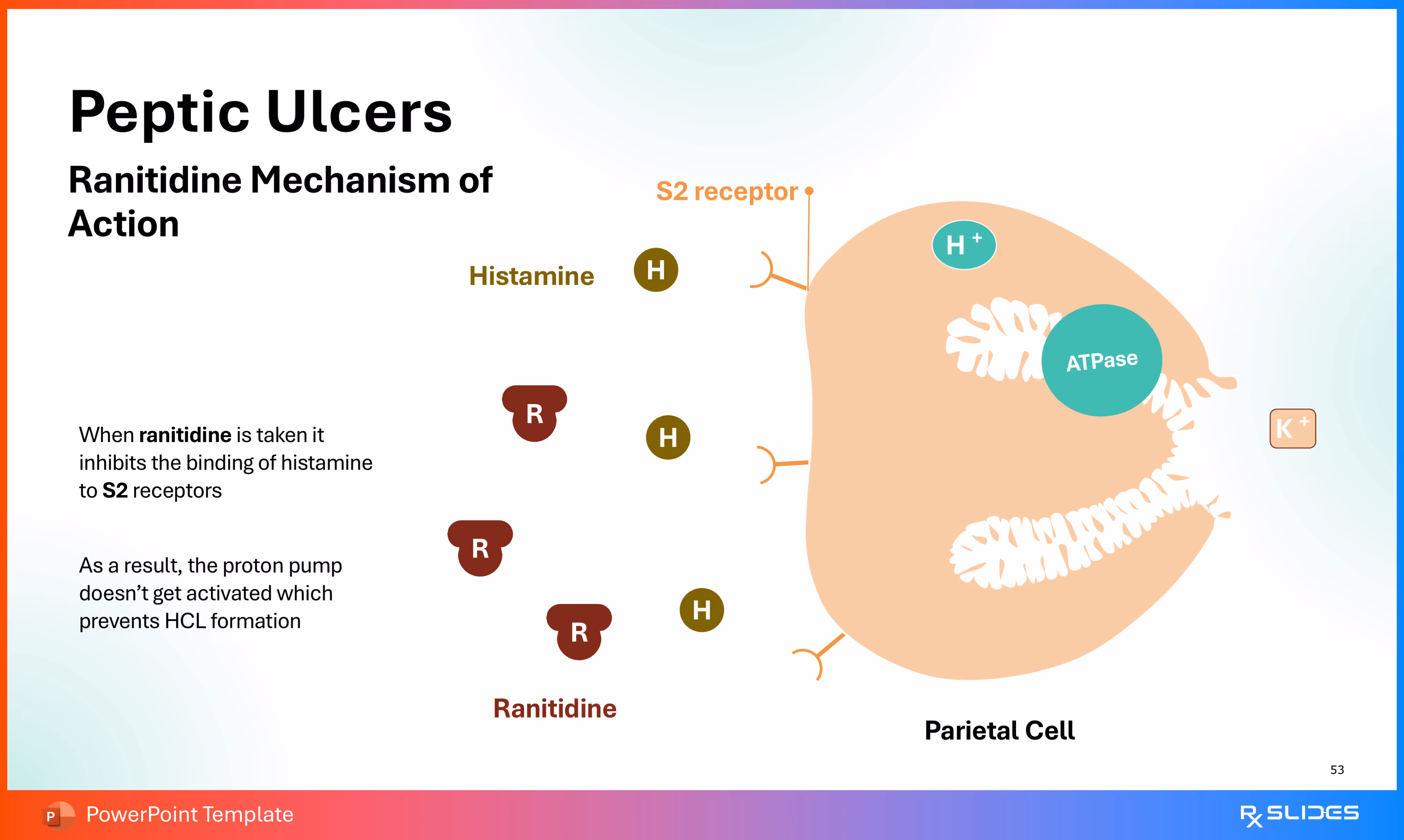
Animation is crucial for demonstrating molecular processes that occur inside cells and at receptor sites, clarifying biochemical steps and signaling pathways.
Templates use animation to illustrate the complete cellular signaling pathway of an adrenergic agonist. The sequence shows:
1. The drug (salmeterol) binding to the receptor.
2. The subsequent activation of the protein.
3. The role of cAMP and the final activation of protein kinase (PK). This entire process visually explains how the drug ultimately leads to the therapeutic effect of widened bronchi.
2. Leukotriene modifiers (represented by small blue blocks) intervene in the area of the bronchiole to counteract the effects that cause mucus secretion and bronchoconstriction.
2. Visualizing Drug-Target Interaction and Inhibition
Animation transforms static diagrams of inhibition into dynamic sequences that show precisely how a drug physically disrupts a process.
Animations are employed to illustrate the normal bacterial action of DNA replication, which ciprofloxacin is designed to disrupt. Subsequent animated slides demonstrate the drug's mechanism, showing:
1. The molecular representation of the drug interfering with the bacterial DNA structure.
2. The drug disrupts the action of associated bacterial enzymes, like DNA gyrase (topoisomerase II) and topoisomerase IV, which are vital for managing DNA supercoiling and separation. The goal is to "visualize the drug's journey as it targets and inhibits DNA replication."
General Benefits of Animated MoA Slides
The use of animation is highlighted as a key feature that maximizes audience engagement and learning:
• Engagement and Retention:
Animated slides capture attention and enhance understanding. They transform passive learning into an active experience and help foster deeper understanding and knowledge retention.
• Clarity and Conciseness:
Animation simplifies the mechanisms of drugs, transforming complex medical concepts into clear and engaging visuals.
• Storytelling for Science:
Animated slides are part of an approach that combines medicine and storytelling to make a lasting impact.


.avif)
.avif)
.avif)