Peptic Ulcer Disease PowerPoint Template
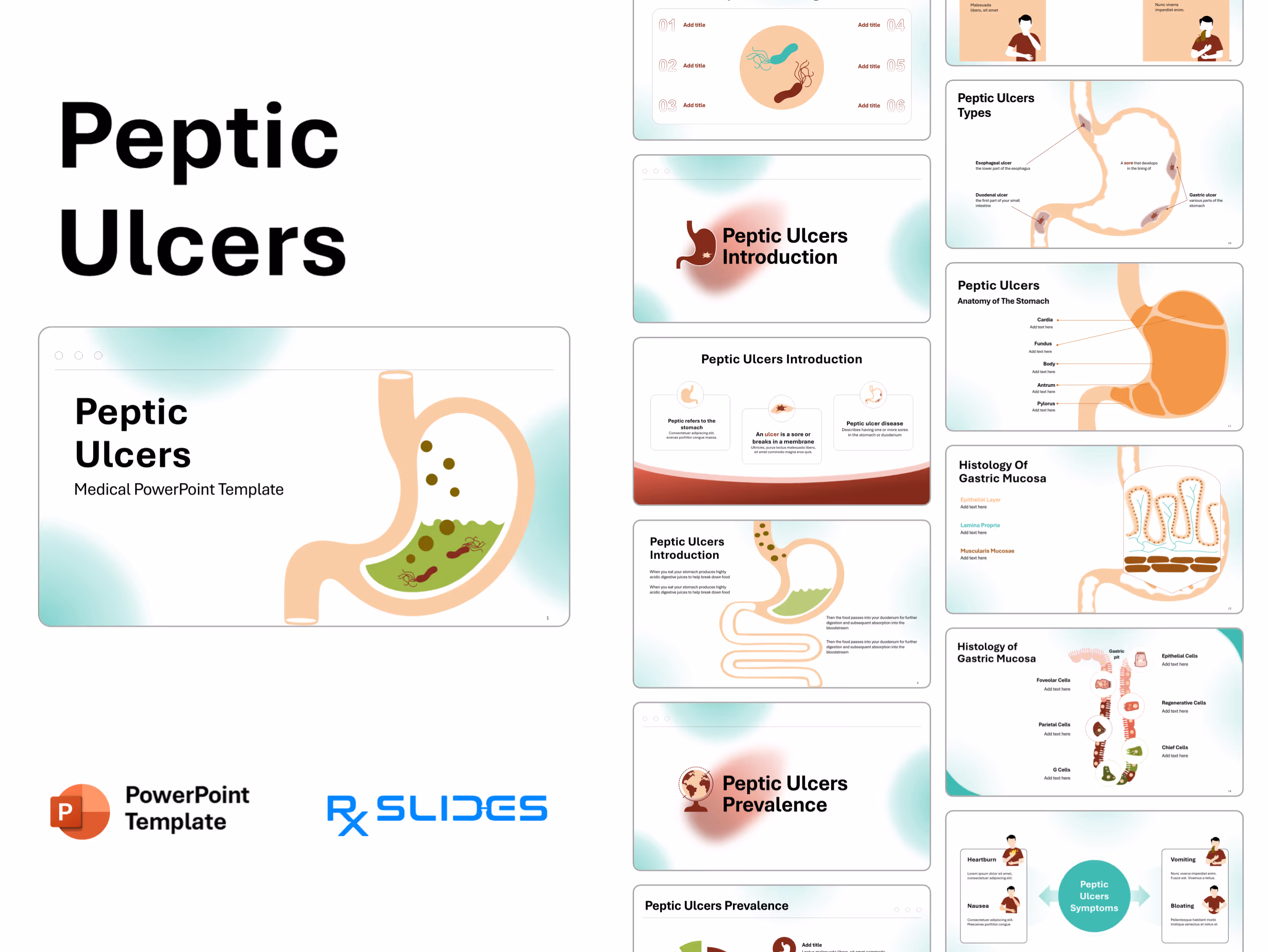
Peptic Ulcer Disease (PUD) Presentation: Medical PowerPoint Template
- The Peptic Ulcer Disease (PUD) PPT template is an animated medical PowerPoint template that will help you realize the full potential of your presentation.
- Our slides, which include medical animations and infographics, will attract your audience.
- You can take advantage of our demonstrated infographics to provide your audience with a dynamic and appealing presentation of Peptic Ulcer Disease (PUD).
- You can notice that the Structure and Function of the Digestive System PowerPoint Templates provides everything you need to create a clear and concise gastrointestinal presentation.
Peptic Ulcer Disease (PUD) PowerPoint Template Preview
Peptic Ulcer Disease (PUD) PowerPoint Template Content
Slide 1 - Title Slide
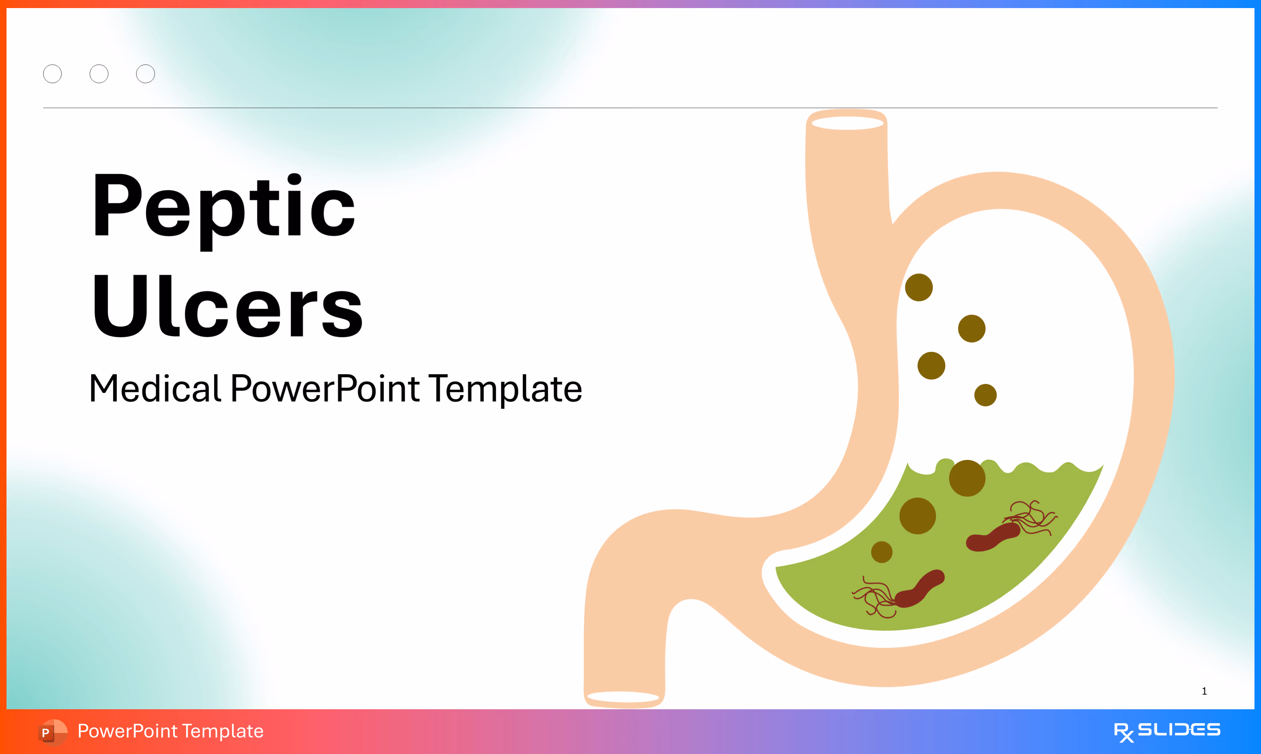
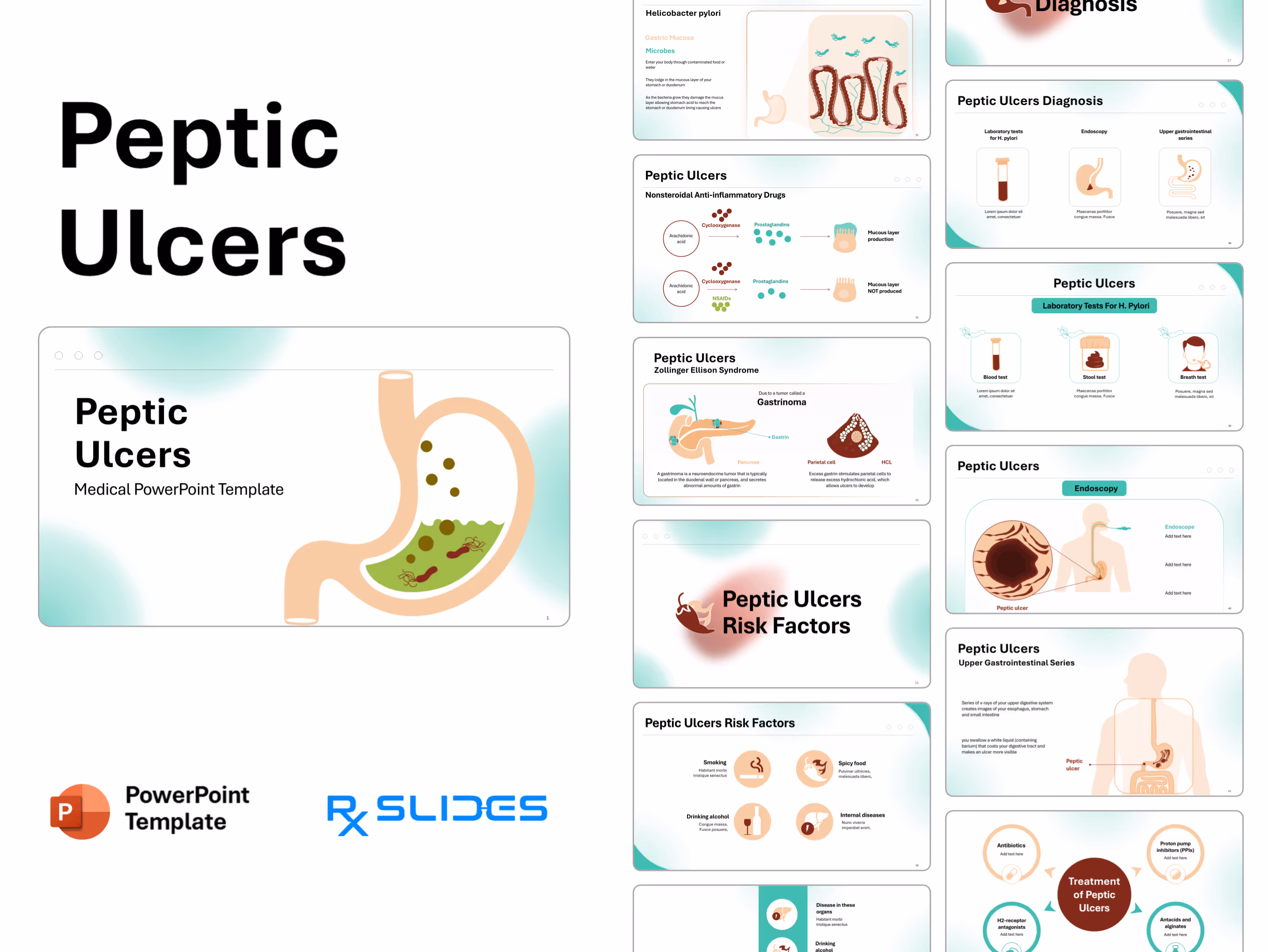
This is the title slide for the new medical template focusing on Peptic Ulcers.
- A stylized illustration of the human stomach is on the right side. Inside the stomach, there is green fluid (stomach acid/contents) with red, helical/rod-shaped bacteria (representing Helicobacter pylori, the primary cause of peptic ulcers).
Slide 2 - Content/Agenda Slide
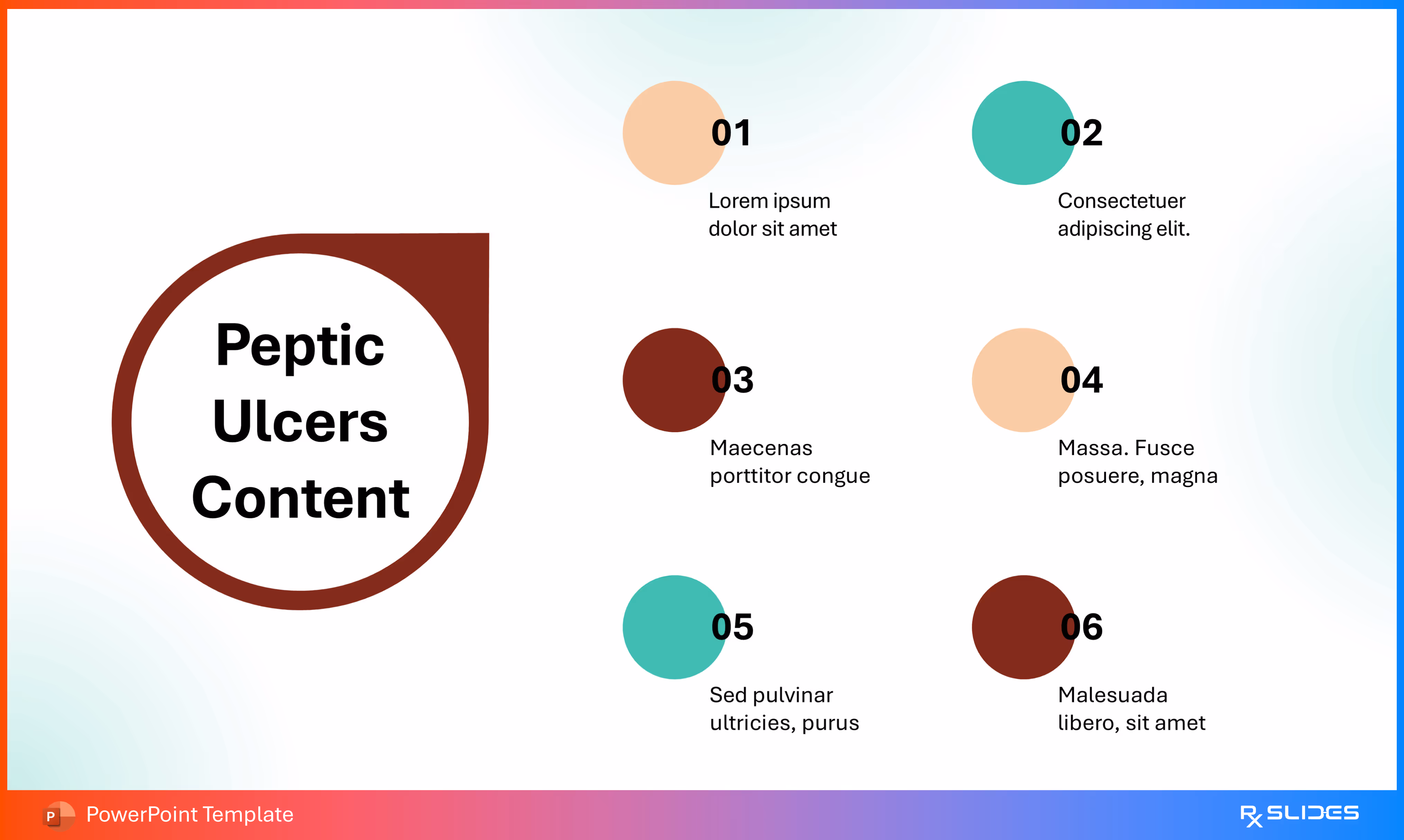
This slide serves as the agenda or table of contents, outlining the six main sections or topics that will be covered in the presentation.
- A large, circular, dark red and light red graphic on the left with the text "Peptic Ulcers Content."
- Six numbered circles, each with a different color (shades of red, beige, and teal) and a placeholder text box, are arranged in two vertical columns to the right of the central element
Slide 3 - Agenda Slide (Alternative Layout)
.avif)
This slide provides an alternative, centralized layout for the presentation agenda, focusing on the microbial aspect of the disease.
- A large, circular graphic in the middle contains two stylized, flagellated bacteria (one teal, one dark red), visually representing Helicobacter pylori.
- Six numbered points (01 through 06) with placeholder text boxes are arranged symmetrically around the central graphic, three on the left and three on the right.
Slide 4 - Peptic Ulcers Introduction (Section Divider)
.avif)
This is a transitional slide introducing the Introduction section of the template.
- Title: Features the text "Peptic Ulcers Introduction".
- Visual Element: A simplified illustration of the human stomach is on the left, colored in dark red/maroon. A small, stylized white ulcer or lesion is clearly visible within the organ's wall.
Slide 5 - Peptic Ulcers Definition
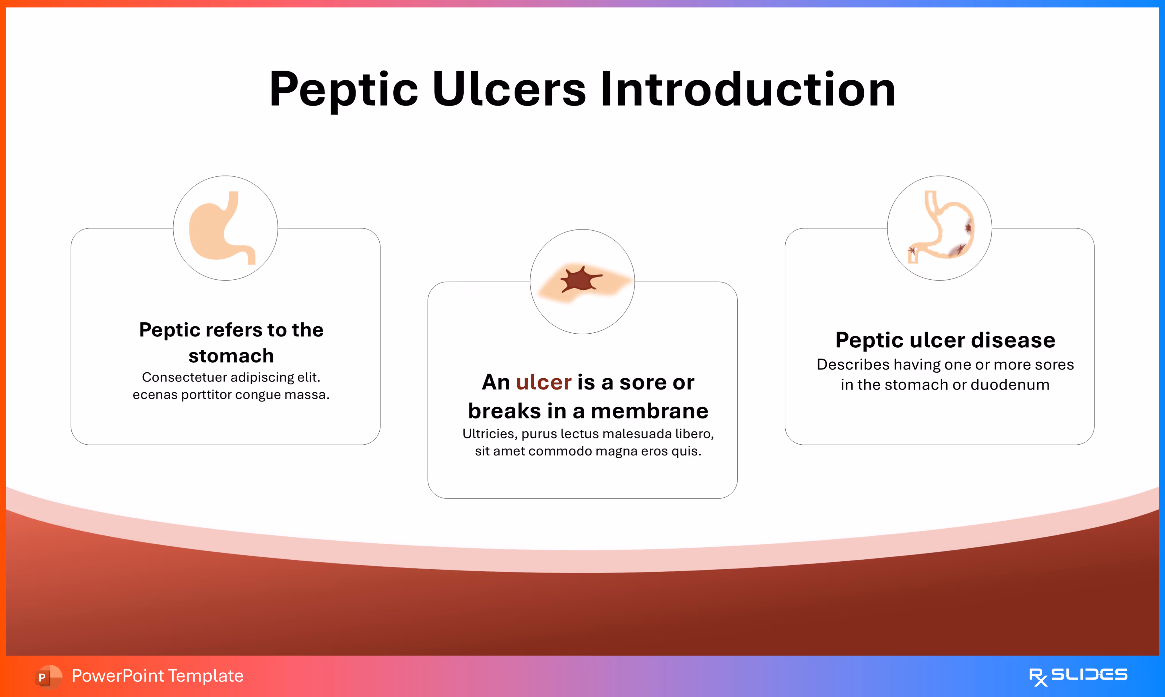
A detailed slide providing the definitions of key terms related to peptic ulcer disease, presented in a three-column layout.
- Peptic refers to the stomach (Icon: Stylized illustration of the stomach).
- An ulcer is a sore or breaks in a membrane (Icon: Close-up illustration of a small, red-brown sore/lesion).
- Peptic ulcer disease Describes having one or more sores in the stomach or duodenum (Icon: Stylized illustration of the stomach and duodenum, with a lesion on the duodenum).
Slide 6 - Introduction to Stomach Function
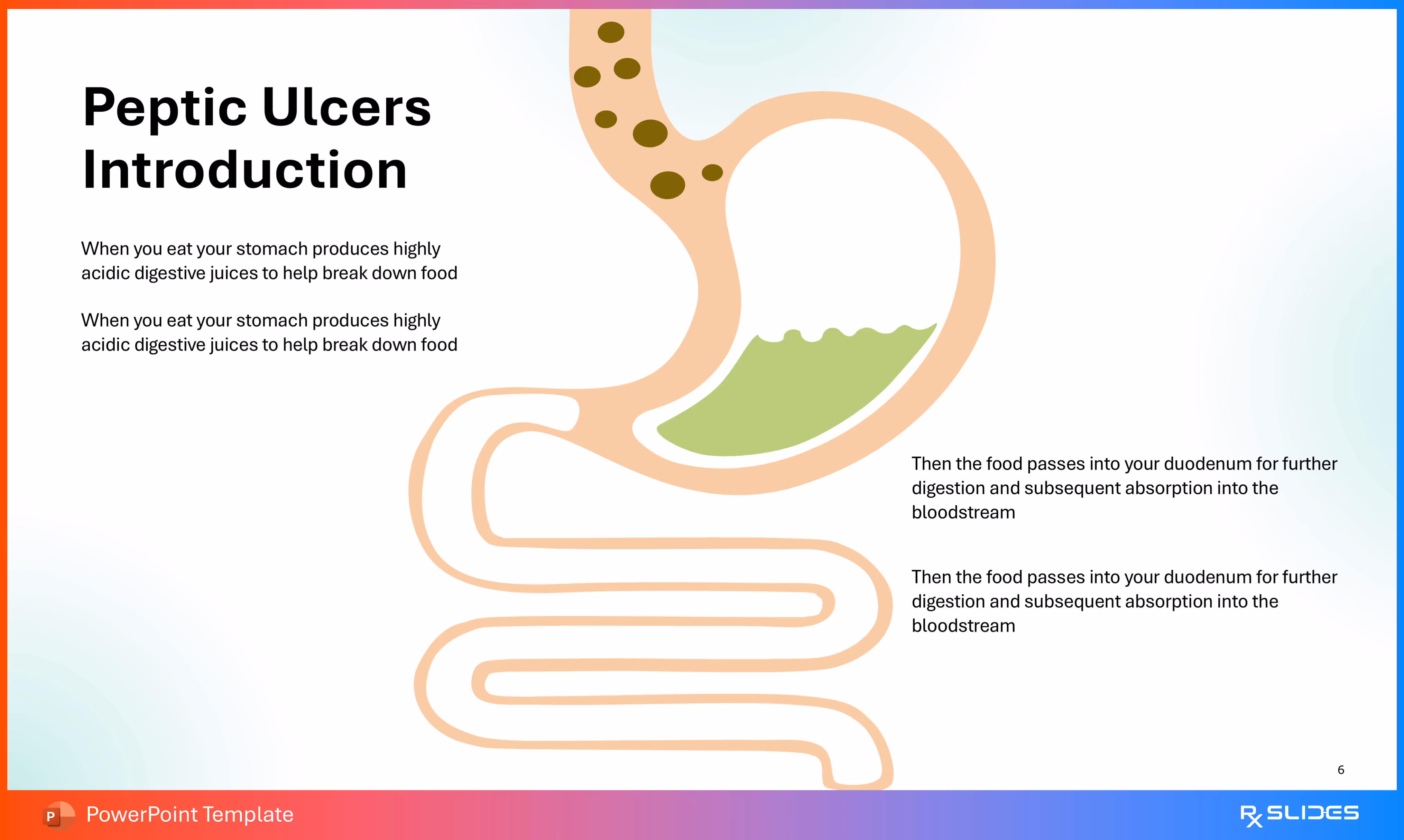
This slide introduces the normal function of the stomach and duodenum as a prelude to explaining how ulcers develop.
The design emphasizes the flow of food and digestive juices.
- A stylized illustration of the stomach leading into the duodenum (the first part of the small intestine). The stomach contains brown food particles and green, acidic fluid.
- Two pairs of repetitive text boxes describe the process:
- Left Side (Stomach Function): "When you eat your stomach produces highly acidic digestive juices to help break down food."
- Right Side (Duodenum Function): "Then the food passes into your duodenum for further digestion and subsequent absorption into the bloodstream."
Slide 7 - Peptic Ulcers Prevalence (Section Divider)
.avif)
- This is a transitional slide introducing the Prevalence (Epidemiology) section of the template.
Slide 8 - Peptic Ulcers Prevalence (Pie Chart Data)
.avif)
A data slide using a pie chart to visually represent the prevalence and distribution of peptic ulcers (likely gastric vs. duodenal) or their causes.
- Dominating the left side, the chart is divided into three sections with approximate percentages, colored in dark red, light peach, and light green:
- Three circular icons on the right, each with a different color and corresponding to the pie chart segments, provide space for explanatory text (e.g., differentiating between gastric ulcer, duodenal ulcer, and other types).
Slide 9 - Peptic Ulcers Types (Section Divider)
.avif)
- This is a transitional slide introducing the Types section of the template, which will likely distinguish between Gastric and Duodenal ulcers.
Slide 10 - Peptic Ulcers Types (Anatomy)
.avif)
A highly illustrative anatomical slide detailing the three main types of peptic ulcers based on their location in the upper gastrointestinal tract.
- A large, stylized illustration of the stomach, esophagus, and duodenum shows the locations of the sores.
- The text defines an ulcer as "A sore that develops in the lining of" the area.
- Arrows point from the definition text to the corresponding lesion in the diagram:
- Esophageal ulcer: Located at the lower part of the esophagus.
- Duodenal ulcer: Located in the first part of your small intestine (duodenum).
- Gastric ulcer: Located in various parts of the stomach.
Slide 11 - Anatomy of The Stomach
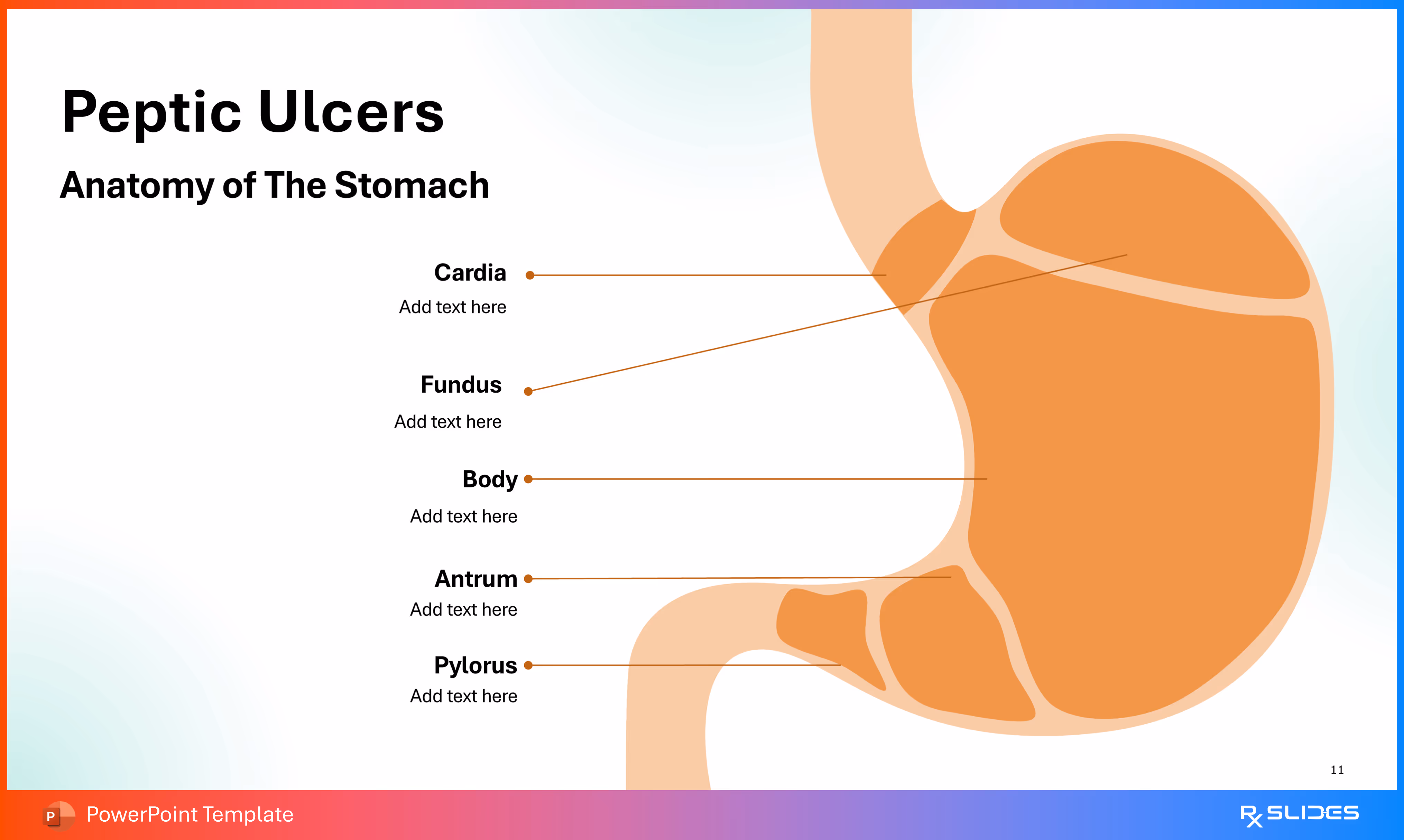
A highly illustrative anatomical slide detailing the five main sections of the stomach. This slide serves to locate where gastric ulcers commonly occur.
- A large, stylized illustration of the stomach is on the right, shown in a solid orange/beige color.
- Text boxes with leader lines point to the different anatomical regions of the stomach:
- Cardia
- Fundus
- Body
- Antrum
- Pylorus
Slide 12 - Anatomy of The Stomach (Alternative Layout)
.avif)
- An alternative, clear anatomical slide detailing the five main sections of the stomach.
Slide 13 - Histology of Gastric Mucosa
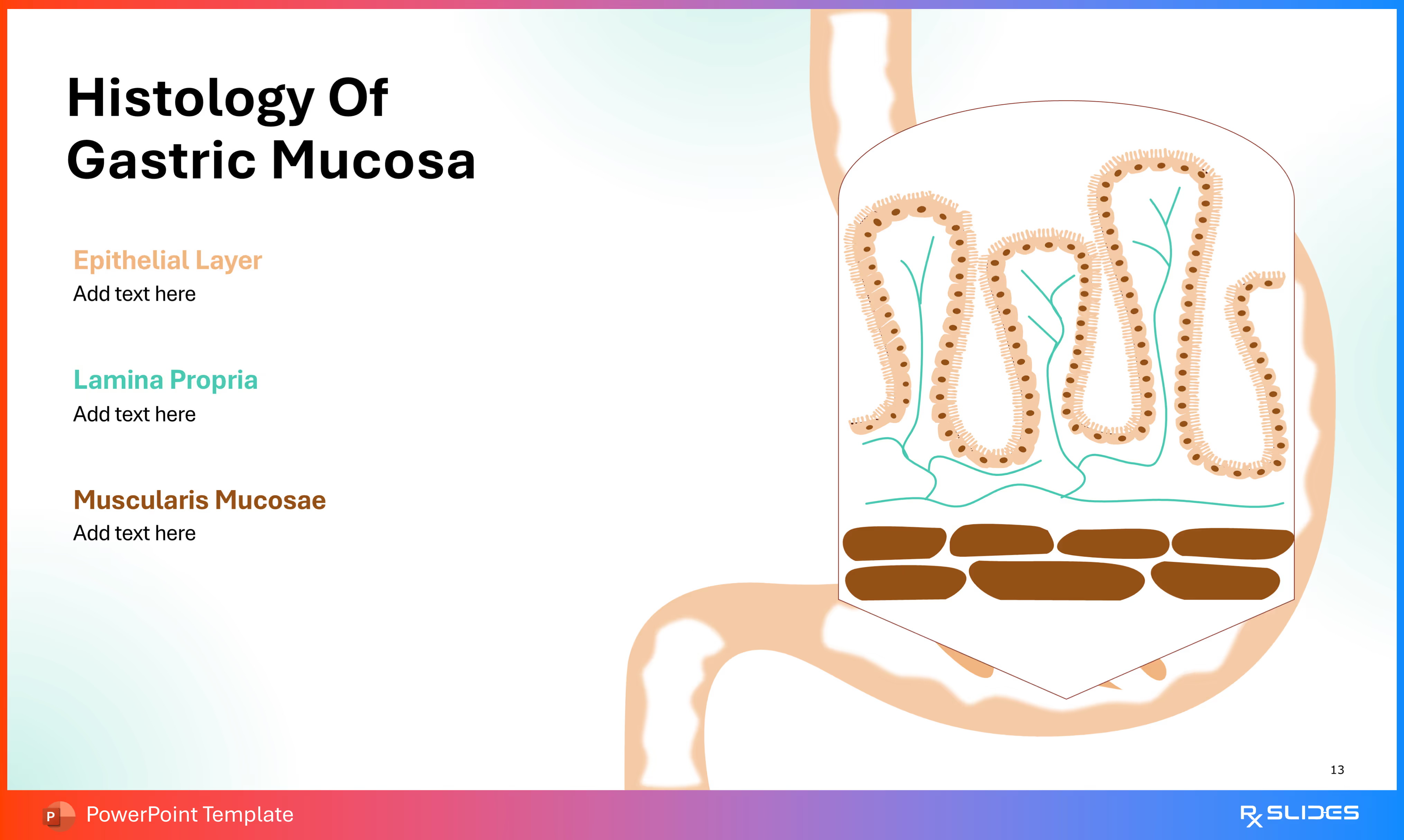
A detailed anatomical diagram focusing on the layers of the stomach wall, crucial for understanding where ulcers form and the protective mechanisms that break down.
- A large, magnified, cross-sectional illustration of the stomach wall layers on the right side.
- The diagram clearly shows the folds (gastric pits/glands) of the mucosa.
- Three distinct layers are identified by color-coded text and leader lines, with space for text entry:
- Epithelial Layer (Orange/Brown text)
- Lamina Propria (Teal text)
- Muscularis Mucosae (Dark Red/Brown text)
Slide 14 - Histology of Gastric Mucosa (Specialized Cells)
.avif)
A highly detailed cellular diagram focusing on the various cell types lining the gastric pit and glands, which are essential for mucus protection and acid production.
- A large, magnified, and labeled cross-sectional illustration of a gastric pit/gland.
- Different cell types are highlighted with small, circular inset icons.
- Text boxes with leader lines point to the corresponding cells, with space for text entry:
- Epithelial Cells
- Foveolar Cells
- Regenerative Cells
- Parietal Cells
- Chief Cells
- G Cells
Slide 15 - Peptic Ulcers Symptoms (Section Divider)
.avif)
This is a transitional slide introducing the Symptoms section of the template.
Slide 16 - Peptic Ulcers Symptoms (Key Indicators)
.avif)
This slide presents a list of four common symptoms of peptic ulcers in a balanced, two-column layout around a central title graphic.
- A teal circle in the center is labeled "Peptic Ulcers Symptoms," acting as the focal point.
- The four symptom panels are:
- Heartburn (Icon: Person holding their chest/upper abdomen).
- Nausea (Icon: Person holding their lower abdomen).
- Vomiting (Icon: Person bent over).
- Bloating (Icon: Person holding their lower abdomen).w
Slide 17 - Peptic Ulcers Symptoms (Vertical List)
.avif)
This slide presents an alternative, vertically aligned layout of the four common symptoms of peptic ulcers, emphasizing a clear, numbered progression.
- The text for each symptom is aligned with its corresponding visual icon of a person depicting the symptom.
Slide 18 - Peptic Ulcers Symptoms (Four-Quadrant Layout)
.avif)
This slide presents an alternative, four-quadrant layout of the four common symptoms of peptic ulcers, with the presentation title centralized.
- Features the large, central text "Peptic Ulcers Symptoms".
- The slide is divided into four equal boxes (two top, two bottom), each featuring a symptom and its corresponding icon
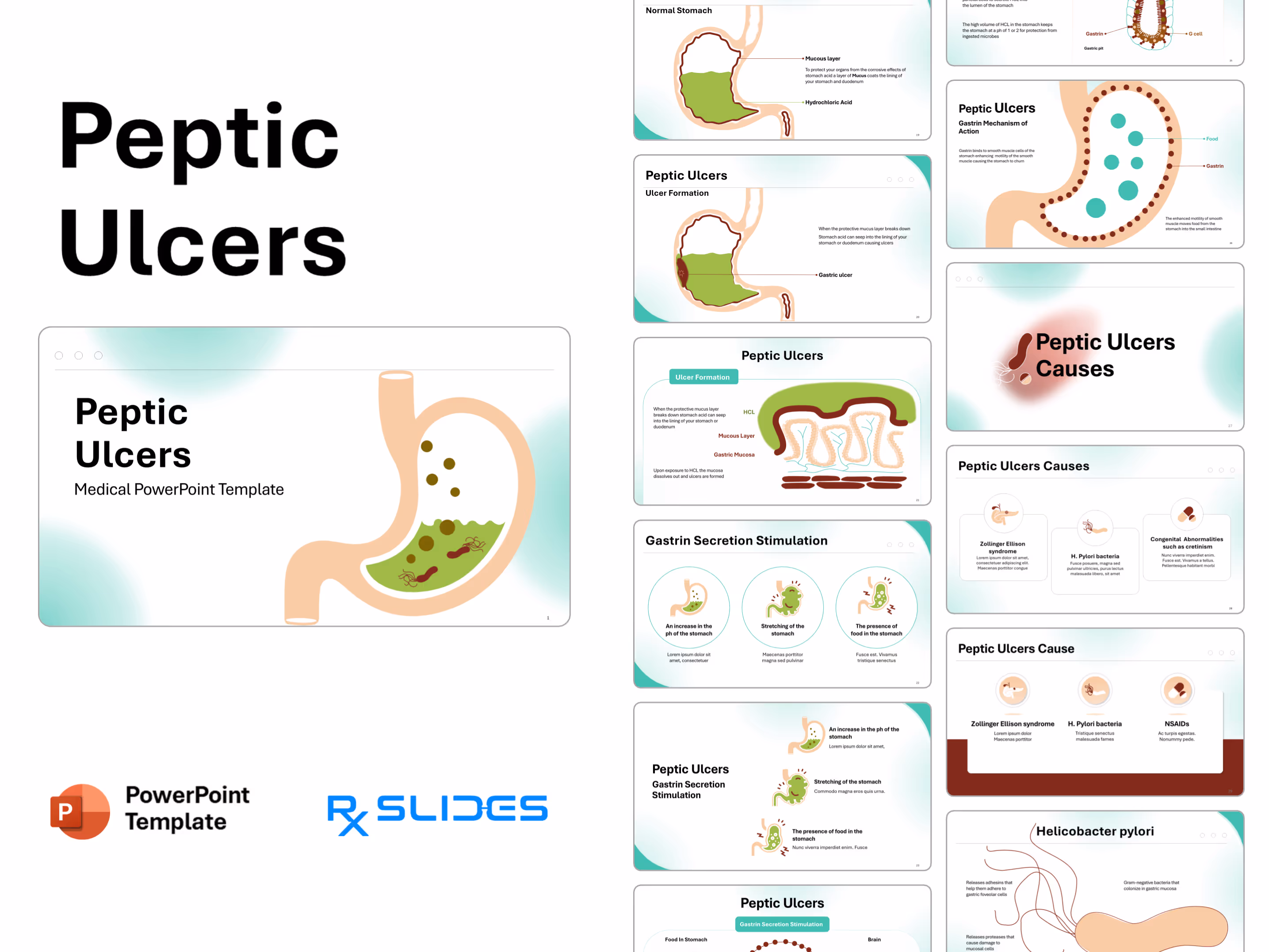
Slide 19 - Normal Stomach (Protective Mechanism)
.avif)
This is an introductory slide to the pathophysiology section, illustrating the normal protective layers of the stomach lining that prevent autodigestion.
- A large, simplified cross-sectional illustration of the stomach and duodenum shows:
- Hydrochloric Acid (Green Fluid): Labeled and filling the lower stomach.
- Mucous Layer (Thick Brown Line): Labeled and lining the inside wall of the stomach.
- The text explains the function of the mucous layer: "To protect your organs from the corrosive effects of stomach acid a layer of Mucus coats the lining of your stomach and duodenum."
Slide 20 - Ulcer Formation (Pathophysiology)
.avif)
This is a core slide in the pathophysiology section, illustrating the breakdown of the stomach's defenses and the resulting ulceration.
- A large, simplified cross-sectional illustration of the stomach and duodenum, similar to Slide 19, but now showing a lesion:
- Gastric ulcer: An arrow points directly to a deep, stylized red lesion with a white core in the stomach wall.
- Hydrochloric Acid (Green Fluid): The fluid is shown seeping into the compromised lining at the site of the ulcer.
- The accompanying text clearly explains the process: "When the protective mucus layer breaks down Stomach acid can seep into the lining of your stomach or duodenum causing ulcers."
Slide 21 - Ulcer Formation (Histological Detail)
.avif)
This is a highly magnified, cross-sectional diagram detailing the process of ulceration at the cellular level of the gastric mucosa.
- A large, detailed illustration of the layers of the stomach wall (similar to Slide 13), showing the breakdown of protection:
- HCL (Green Layer): The hydrochloric acid is shown resting on the compromised Mucous Layer (thick brown line).
- Gastric Mucosa (Pale, folded layer): The folds are visible beneath the mucous layer.
- Two text boxes explain the mechanism:
- "When the protective mucous layer breaks down stomach acid can seep into the lining of your stomach or duodenum."
- "Upon exposure to HCL the mucosa dissolves out and ulcers are formed."
Slide 22 - Gastrin Secretion Stimulation
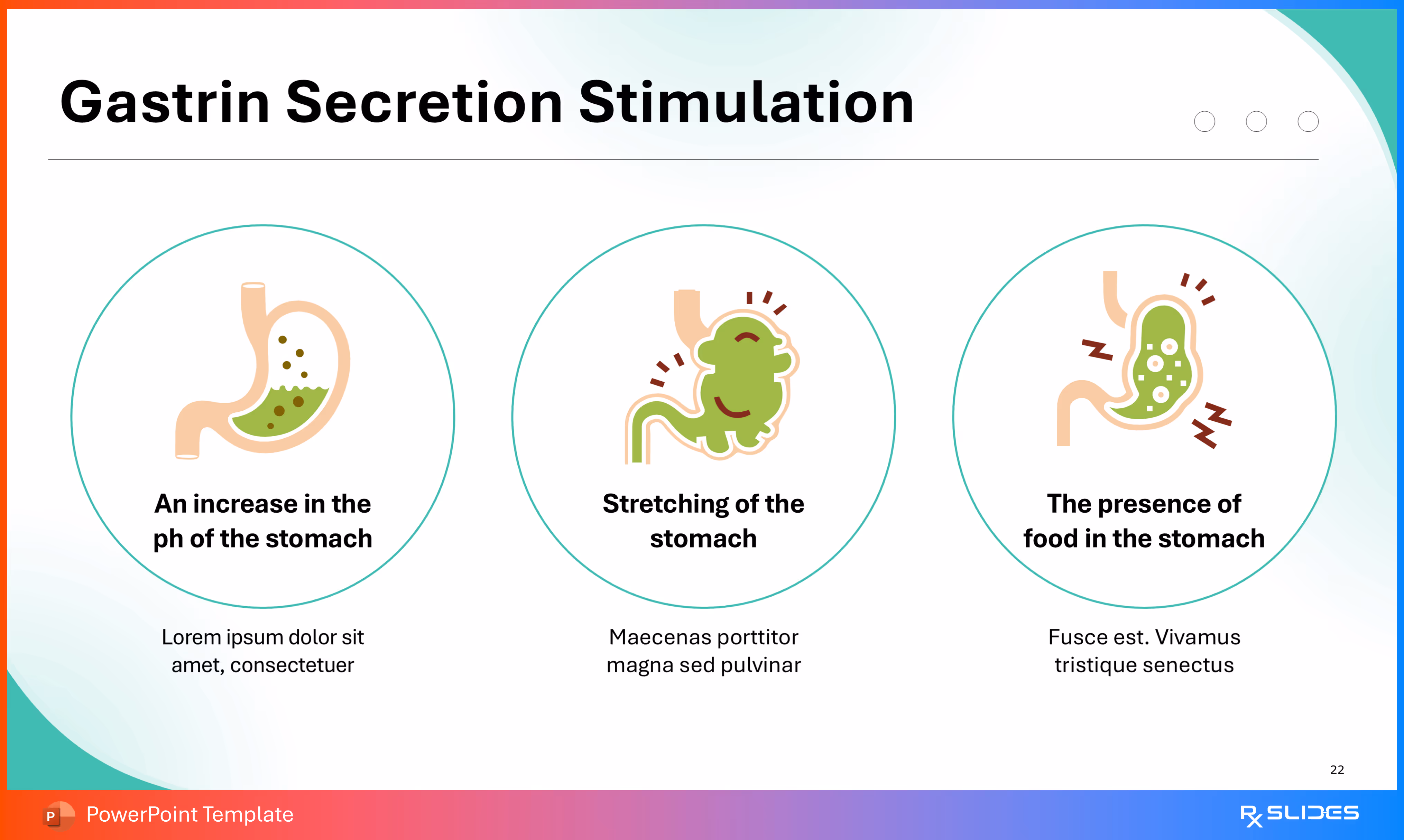
This slide uses a three-point circular layout to illustrate the three main physiological stimuli that lead to the secretion of Gastrin, which then promotes hydrochloric acid (HCL) release and contributes to ulcer formation.
- Each point is represented by a large circular icon featuring a stylized stomach illustration, defining a cause for Gastrin release:
- An increase in the pH of the stomach (Icon: Stomach with green liquid and brown particles, indicating a change in pH).
- Stretching of the stomach (Icon: Enlarged, green stomach with Z or spark lines, representing nerve signals from distension).
- The presence of food in the stomach (Icon: Stomach with green liquid and particles, with nervous system Z lines, representing chemical presence).
Slide 23 - Gastrin Secretion Stimulation (Vertical Layout)
.avif)
This slide provides an alternative, stacked layout to illustrate the three main physiological stimuli that trigger the secretion of Gastrin, which increases acid production and contributes to ulcer formation.
- The three points are arranged vertically down the right side, each featuring a stylized stomach illustration and defining a cause for Gastrin release
Slide 24 - Gastrin Secretion Pathway (Neural Mechanism)
.avif)
This is a detailed physiological slide illustrating the neural (vagal) pathway that controls Gastrin secretion, which ultimately leads to acid production.
- The slide is divided into three parts:
- Left (Input): Food In Stomach (illustrated by the stomach with contents) and Gastrin Secretion from G cells (illustrated by a magnified inset of G-cells).
- Center (Pathway): A circular pathway made of brown dots represents the vagal signaling. The text explains the action: "Vagal signaling induced by the parasympathetic nervous system to stimulate the release of gastrin from g cells."
- Right (Control): An illustration of the Brain and Medulla, representing the central nervous system control of the parasympathetic (vagal) signals.
Slide 25 - Gastrin Mechanism of Action
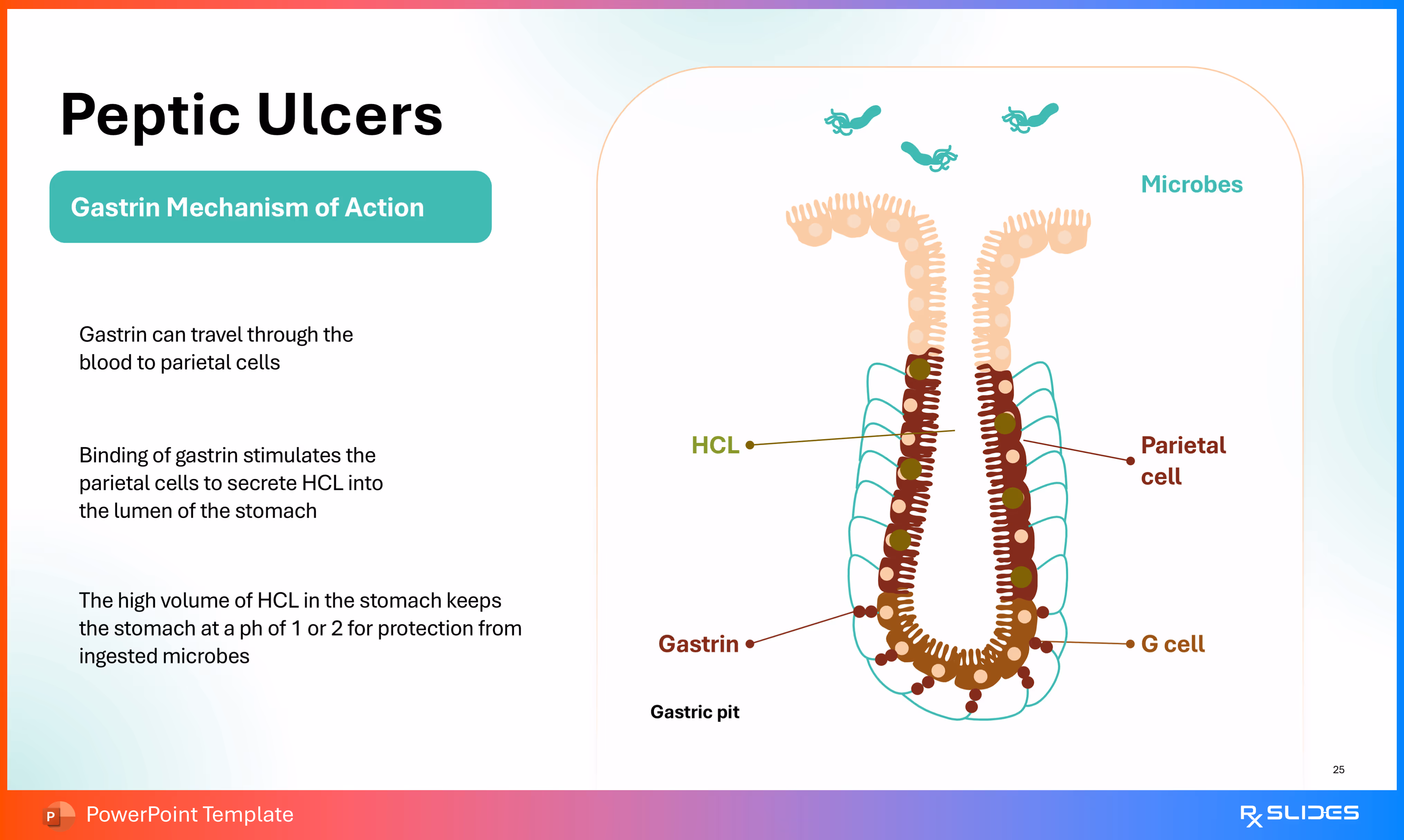
This is a detailed biochemical and histological slide illustrating the molecular mechanism by which Gastrin stimulates the production of hydrochloric acid (HCL), a key factor in ulcer development.
- A highly magnified illustration of a gastric pit/gland showing the cellular interaction:
- G cell: Located at the bottom of the gland, releasing Gastrin.
- Parietal cell: Targeted by Gastrin, responsible for secreting HCL into the lumen (top).
- Microbes: Stylized bacteria are shown above the gland, indicating the protective role of acid.
- Three bullet points explain the flow of the mechanism:
- Gastrin can travel through the blood to parietal cells (hormonal action).
- Binding of gastrin stimulates the parietal cells to secrete HCL into the lumen of the stomach (the effect).
- The high volume of HCL in the stomach keeps the stomach at a pH of 1 or 2 for protection from ingested microbes (the purpose of the acid).
Slide 26 - Gastrin Mechanism of Action (Motility)
.avif)
This is a physiological slide illustrating the second key function of Gastrin: enhancing the motility and churning action of the stomach, which aids in digestion and emptying.
- A simplified illustration of the stomach and duodenum is shown.
- The wall of the stomach is lined with brown dots representing the smooth muscle cells that respond to Gastrin. Teal circles inside the stomach represent Food.
- Text boxes describe the action:
- Left: "Gastrin binds to smooth muscle cells of the stomach enhancing motility of the smooth muscle causing the stomach to churn."
- Right (pointing to Gastrin dot-line): "Gastrin"
- Bottom Right: "The enhanced motility of smooth muscle moves food from the stomach into the small intestine."
Slide 27 - Peptic Ulcers Causes (Section Divider)
.avif)
This is a transitional slide introducing the Causes section of the template.
- A prominent illustration of the Helicobacter pylori bacterium, shown as a stylized, flagellated, dark red/brown rod.
- This visually cues the primary etiological agent of peptic ulcer disease.
Slide 28 - Peptic Ulcers Causes (Three Main Etiologies)
.avif)
This slide presents a three-column layout detailing the three primary causes of peptic ulcers.
- The slide features three distinct panels, each with an icon representing a cause:
- Zollinger-Ellison syndrome: Icon of the pancreas/duodenum with a small tumor-like growth, representing the tumor that secretes high levels of Gastrin.
- H. Pylori bacteria: Icon of a flagellated bacterium, the most common cause.
- Congenital Abnormalities such as cretinism: Icon of two pills/capsules, representing drug-related causes (e.g., NSAIDs, though the text mentions congenital abnormalities, the drug icon is a common stand-in for non-infectious causes).
- Each panel includes the name of the cause and space for explanatory text:
- Zollinger-Ellison syndrome
- H. Pylori bacteria
- Congenital Abnormalities such as cretinism
Slide 29 - Peptic Ulcers Causes (Three Main Etiologies - Alternative)
.avif)
This slide presents an alternative three-column layout detailing the three most common and specific causes of peptic ulcers.
- Visual Centerpiece: The slide features three distinct panels, each with an icon representing a cause
Slide 30 - Helicobacter pylori (Mechanism of Action)
.avif)
This slide provides a highly detailed, magnified illustration and mechanism of action for Helicobacter pylori (H. pylori), the Gram-negative bacteria that is the main cause of peptic ulcers.
- A large, stylized illustration of the H. pylori bacterium, characterized by its curved rod shape (or spirochete-like appearance) and multiple flagella (whip-like tails).
- Three key mechanisms or facts related to the bacterium are detailed around the illustration:
- Gram-negative bacteria that colonize in gastric mucosa (Defining characteristic and location).
- Releases adhesins that help them adhere to gastric foveolar cells (Mechanism for colonization/attachment).
- Releases proteases that cause damage to mucosal cells (Mechanism for causing the ulceration).
Slide 31 - Helicobacter pylori (Infection Mechanism)
.avif)
This slide visually and textually details the infectious process of H. pylori, showing how the bacteria colonize the gastric mucosa and cause damage.
- A large, highly magnified, cross-sectional illustration of the Gastric Mucosa (the stomach lining folds). Teal, flagellated microbes (H. pylori) are shown:
- Floating above the folds (representing entry/lumen).
- Lodging within and damaging the brown, protective mucous layer.
- Three steps explain the infection process:
- Microbes: "Enter your body through contaminated food or water."
- Colonization: "They lodge in the mucous layer of your stomach or duodenum."
- Damage: "As the bacteria grow they damage the mucus layer allowing stomach acid to reach the stomach or duodenum lining causing ulcers."
Slide 32 - NSAIDs Mechanism (Inhibition of Mucus Production)
.avif)
This is a detailed biochemical slide illustrating the mechanism by which Nonsteroidal Anti-inflammatory Drugs (NSAIDs) inhibit the production of the protective mucous layer, leading to ulceration.
- The slide uses a two-row process diagram comparing the normal pathway to the NSAID-inhibited pathway:
- Top Row (Normal Pathway): Arachidonic acid → Cyclooxygenase (brown dots) → Prostaglandins (teal circles) → Mucous layer production (cell with mucus lining). This shows Prostaglandins are necessary for mucus production.
- Bottom Row (NSAID Effect): Arachidonic acid → Cyclooxygenase (brown dots) is inhibited by NSAIDs (green dots) → Prostaglandins (teal circles are present, but their action is implied to be blocked or production is reduced) → Mucous layer NOT produced (cell lacks mucus lining). This illustrates that NSAIDs interfere with Prostaglandin's protective role, which is key to maintaining the mucous barrier.
Slide 33 - Zollinger-Ellison Syndrome
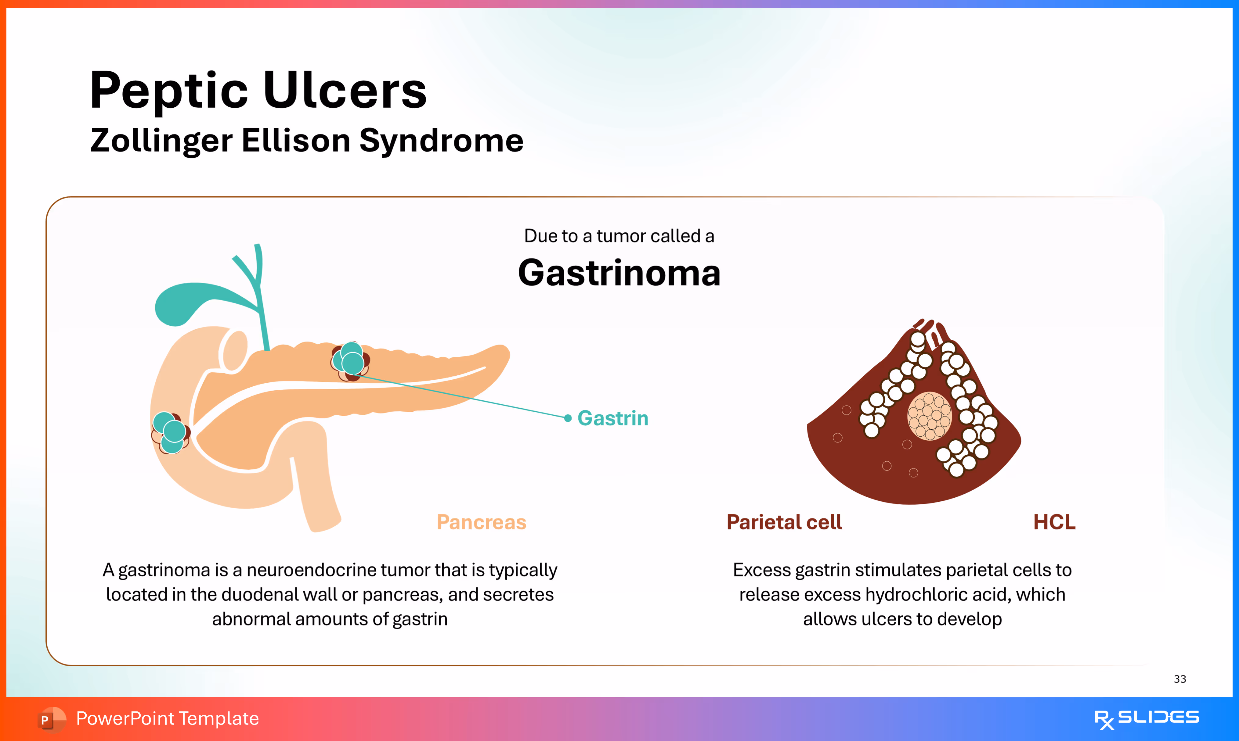
This slide provides an illustration and explanation of Zollinger-Ellison Syndrome (ZES), focusing on how a Gastrinoma tumor leads to excessive acid production and ulceration.
- An illustration of the pancreas and duodenal wall. Two Gastrinoma tumors (teal circles with red borders) are shown on the pancreas and duodenum, releasing Gastrin (teal arrow).
- Text (Bottom Left): Defines the condition: "A gastrinoma is a neuroendocrine tumor that is typically located in the duodenal wall or pancreas, and secretes abnormal amounts of gastrin."
- A magnified, stylized image of a Parietal cell cluster (dark red) with an illustration of its effect.
- Text (Bottom Right): Explains the effect: "Excess gastrin stimulates parietal cells to release excess hydrochloric acid, which allows ulcers to develop." The labels indicate the Parietal cell and the resulting HCL.
Slide 34 - Peptic Ulcers Risk Factors (Section Divider)
.avif)
- This is a transitional slide introducing the Risk Factors section of the template, which often covers lifestyle and environmental contributors.
Slide 35 - Peptic Ulcers Risk Factors (Four Factors)
.avif)
This slide presents the four most common lifestyle and underlying health risk factors for developing peptic ulcers in a four-quadrant, circular icon layout.
- The slide features four large, circular icons, each representing a risk factor:
- Smoking (Icon: Stylized cigarette with smoke, representing the habit).
- Spicy food (Icon: Stylized chili pepper, representing dietary irritants).
- Drinking alcohol (Icon: Stylized wine bottle and glass).
- Internal diseases (Icon: A body organ, likely the liver, with a lightning bolt symbol, representing chronic underlying health conditions that can stress the digestive system or require high NSAID use).
Slide 36 - Peptic Ulcers Risk Factors (Vertical List)
.avif)
This slide presents an alternative, vertical list of four risk factors, separated by a distinct colored vertical bar.
- The four factors are aligned vertically on the right, within a white-teal-maroon vertical strip, each with a corresponding circular icon
Slide 37 - Peptic Ulcers Diagnosis (Section Divider)
.avif)
- This is a transitional slide introducing the Diagnosis section of the template, which will likely cover testing methods for ulcers and H. pylori.
Slide 38 - Peptic Ulcers Diagnosis (Tests)
.avif)
This slide presents a three-column layout detailing the three primary methods used to diagnose peptic ulcers and their common underlying cause (H. pylori).
- The slide features three distinct panels with corresponding icons:
- Laboratory tests for H. pylori (Icon: A test tube filled with blood, representing blood, stool, or breath tests).
- Endoscopy (Icon: A stylized stomach showing an endoscope or a visual representation of the procedure used to directly view the ulcer).
- Upper gastrointestinal series (Icon: A stomach and upper small intestine with small dots, representing the movement of contrast material used in imaging).
Slide 39 - Laboratory Tests for H. pylori
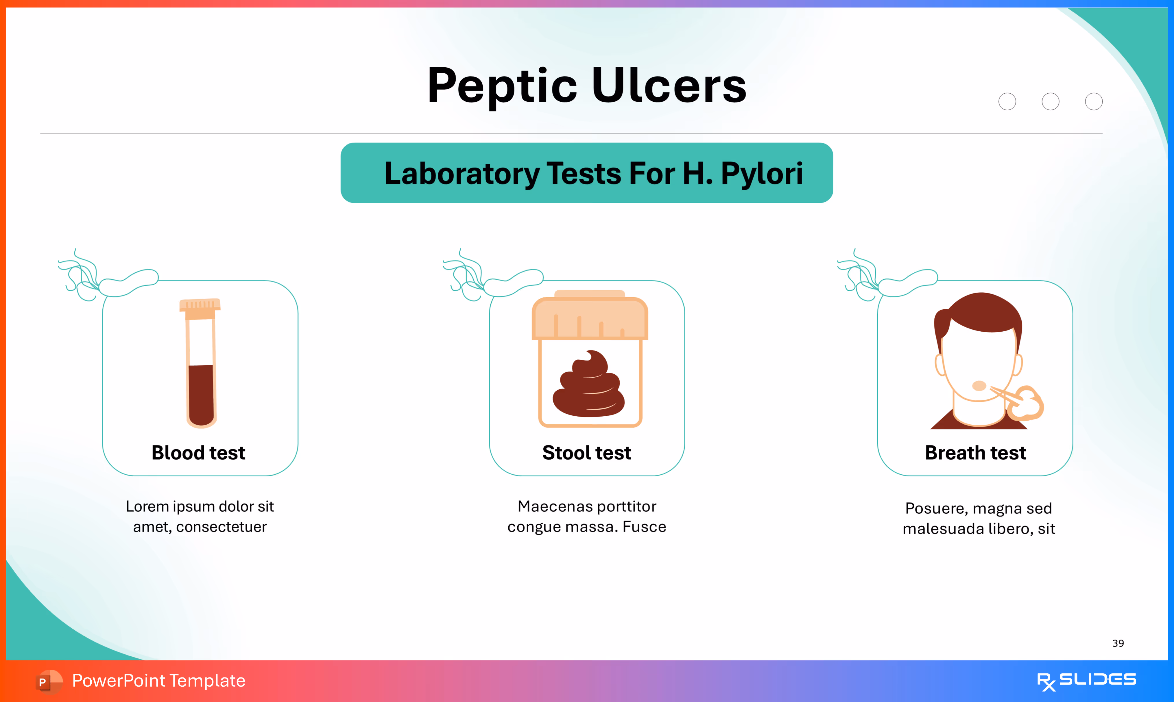
This slide provides specific details on the three most common non-invasive laboratory tests used to diagnose the presence of H. pylori infection.
- The slide features three distinct panels with corresponding icons in a three-column layout:
- Blood test (Icon: A test tube filled with blood).
- Stool test (Icon: A specimen container with a stylized stool sample).
- Breath test (Icon: A man exhaling into a specialized mouthpiece/device).
Slide 40 - Endoscopy
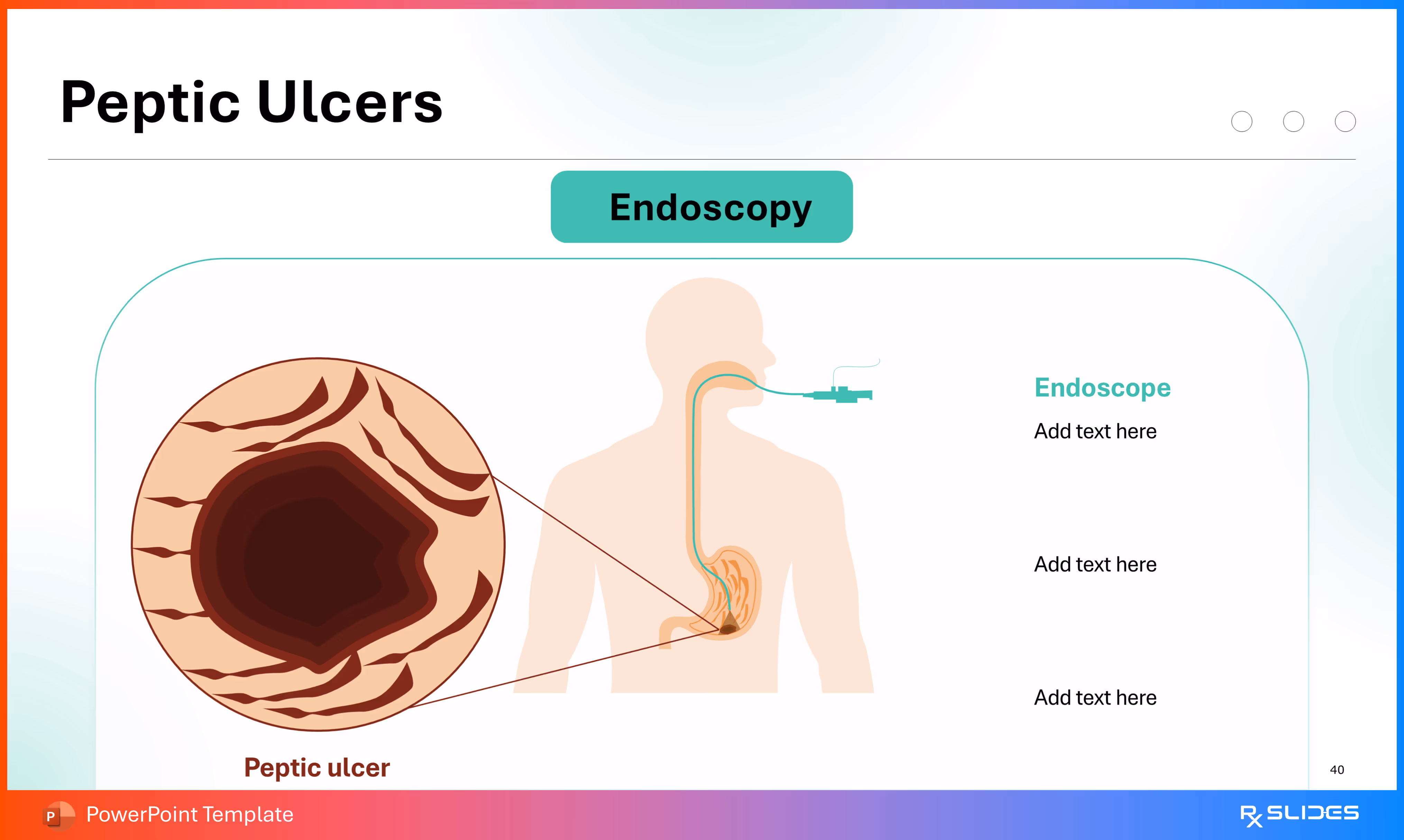
This slide provides a detailed visual explanation of the Endoscopy procedure, which is the gold standard for directly diagnosing peptic ulcers.
- The slide contains two key illustrations:
- Procedure Diagram (Right): A diagram showing the outline of a human torso and upper GI tract. An endoscope (a thin, flexible tube) is shown being passed down the esophagus and into the stomach.
- Magnified Ulcer View (Left): A large, magnified circular inset shows a close-up of a severe peptic ulcer inside the stomach lining. The lesion is dark, deep, and surrounded by inflamed tissue.
- Text placeholders are available on the right side to describe the procedure, the view, and any subsequent actions (like biopsy or treatment).
Slide 41 - Upper Gastrointestinal Series

This slide provides a visual and textual explanation of the Upper Gastrointestinal (GI) Series, an imaging technique used to diagnose peptic ulcers.
- A large, stylized diagram of a human torso showing the upper digestive system: the esophagus, stomach, and small intestine.
- A peptic ulcer (dark red lesion) is visibly marked on the duodenum/stomach boundary.
- The text explains the procedure:
- "Series of X-rays of your upper digestive system creates images of your esophagus, stomach and small intestine".
- "You swallow a white liquid (containing barium) that coats your digestive tract and makes an ulcer more visible".
Slide 42 - Peptic Ulcers Treatment (Section Divider)
.avif)
- This is a transitional slide introducing the Treatment section of the template, which will likely detail the various pharmacological and lifestyle interventions for peptic ulcer disease.
Slide 43 - Treatment of Peptic Ulcers (Pharmacology)
.avif)
This slide presents a four-point circular diagram that organizes the four main classes of medication used to treat peptic ulcer disease, centered around a main theme.
- A large, dark red circle in the center is labeled "Treatment of Peptic Ulcers".
- Four surrounding circular icons (connected to the center by arrows) detail the medication types:
- Antibiotics (Top Left, Icon: Capsule/pill): Used primarily to eradicate H. pylori infection.
- Proton pump inhibitors (PPIs) (Top Right, Icon: Pill): Used to strongly suppress stomach acid production.
- Antacids and alginates (Bottom Right, Icon: Medicine bottle): Used for immediate, short-term relief of symptoms by neutralizing acid.
- H2-receptor antagonists (Bottom Left, Icon: Two pills): Used to reduce acid production by blocking histamine receptors on parietal cells.
Slide 44 - Treatment of Peptic Ulcers (Vertical List)
.avif)
- This slide presents a clear, vertical list detailing the four main classes of medication used to treat peptic ulcer disease, radiating from a central theme circle.
Slide 45 - Treatment of Peptic Ulcers (Four-Quadrant Flow)
.avif)
- This slide presents an alternative, numbered flow diagram detailing the four main pharmacological treatments for peptic ulcers in a circular, four-quadrant layout.
Slide 46 - Peptic Ulcers Antibiotics (Triple Therapy)
.avif)
This slide provides specific details on the antibiotic-based treatment for H. pylori-induced peptic ulcers, emphasizing the common Triple Therapy regimen.
- Two Panels (Triple Therapy):
- Left Panel (Formula): Features a stylized medicine bottle outline containing the components of the triple therapy regimen:
- PPI
- Clarithromycin
- Amoxicillin
- Right Panel (Visual/Name): Features a large green circle and a pill pack, labeled "Clarithromycin Triple Therapy".
- Left Panel (Formula): Features a stylized medicine bottle outline containing the components of the triple therapy regimen:
Slide 47 - Antibiotics Mechanism of Action (on H. Pylori)
.avif)
This slide provides a large, magnified illustration of the stomach lining to show how Antibiotics act to eradicate the H. pylori bacteria, a key step in ulcer healing.
- A large, cross-sectional view of the stomach lining (mucosal folds).
- H. Pylori (brown, flagellated rods) are shown lodged in the mucous layer (light green/yellow layer).
- Antibiotic pills (small green circles and capsules) are shown entering the stomach and targeting the H. Pylori.
- Two text placeholders are provided on the left to detail the specific action of the antibiotics, such as preventing bacterial growth or killing the organisms.
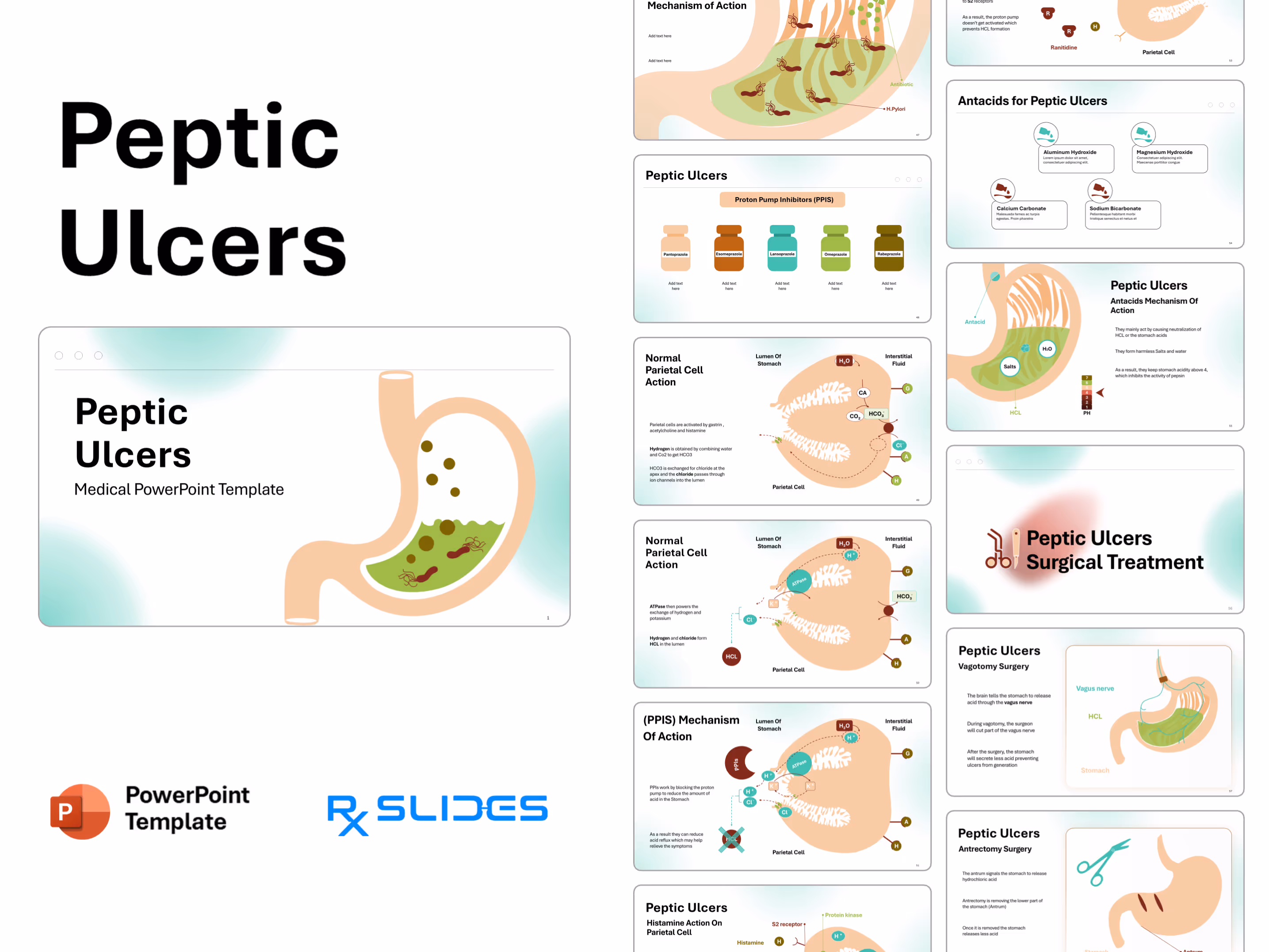
Slide 48 - Proton Pump Inhibitors (PPIs)
.avif)
This slide details the five most common generic examples of Proton Pump Inhibitors (PPIs), a major drug class used in peptic ulcer treatment to suppress acid production.
- The slide shows five stylized medicine bottles of varying colors, each labeled with a specific PPI drug name:
- Pantoprazole (Light beige bottle).
- Esomeprazole (Brown bottle).
- Lansoprazole (Teal bottle).
- Omeprazole (Lime green bottle).
- Rabeprazole (Dark olive bottle).
- This slide functions as a visual catalog of the major drugs in the PPI class.
Slide 49 - Normal Parietal Cell Action
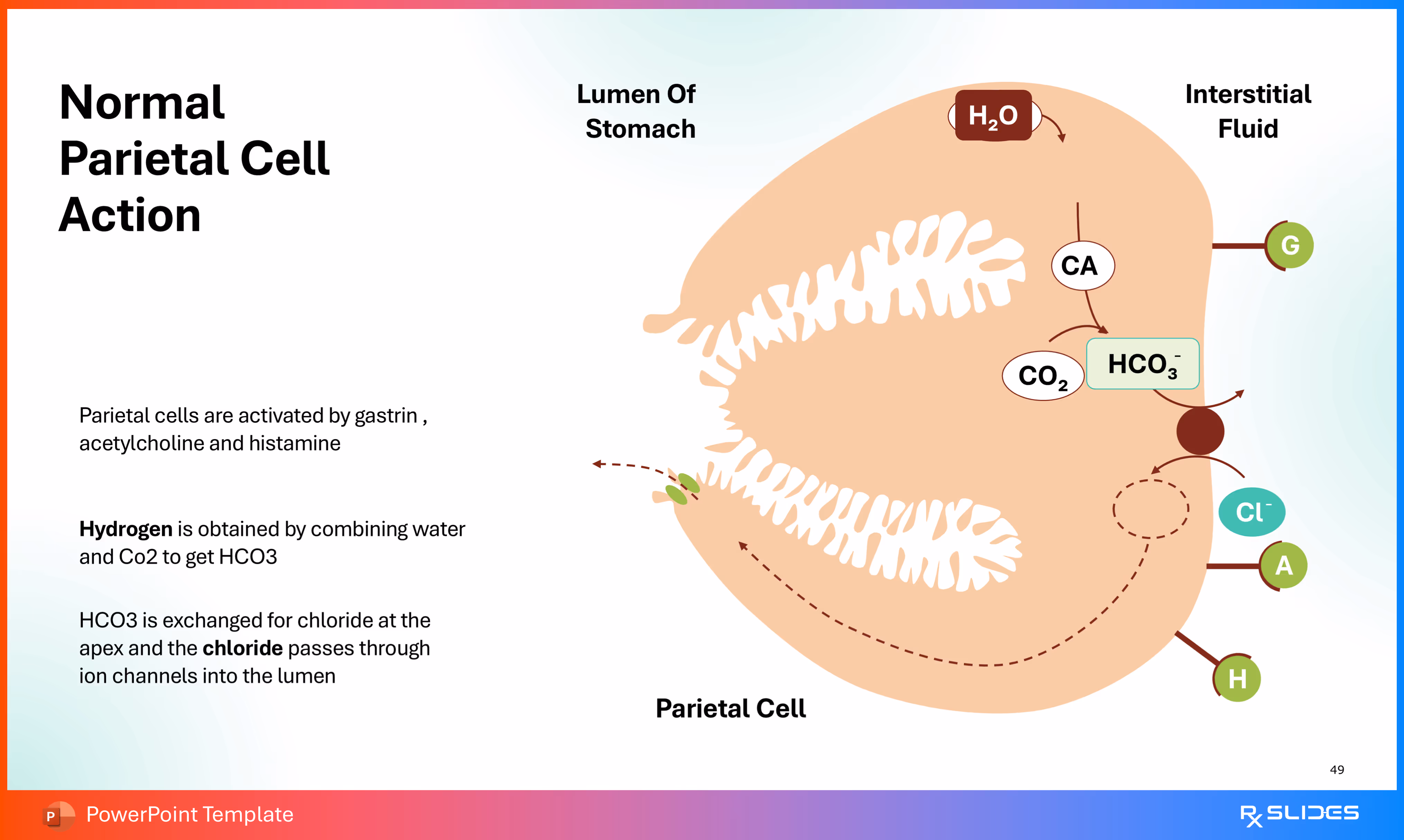
This slide provides a highly detailed, magnified diagram illustrating the normal biochemical mechanism of Hydrochloric Acid (HCL) secretion by the Parietal Cell, the process that PPIs inhibit.
- A large, stylized illustration of a Parietal Cell showing the internal machinery and channels.
- It illustrates the roles of Gastrin (G), Acetylcholine (A), and Histamine (H) in activation.
- The diagram shows the conversion of H2O and CO2 to HCO3− (bicarbonate) and Hydrogen (H+) via Carbonic Anhydrase (CA).
- It shows the exchange of HCO3− for Chloride (Cl−), and the subsequent secretion of Cl− and H+ into the Lumen of the Stomach.
- Key Text:
- Parietal cells are activated by gastrin, acetylcholine, and histamine.
- Hydrogen is obtained by combining water and CO2 to get HCO3−.
- HCO3− is exchanged for chloride at the apex and the chloride passes through ion channels into the lumen.
Slide 50 - Normal Parietal Cell Action (Secretion Phase)
.avif)
This slide continues the detailed, magnified diagram of the Parietal Cell, focusing on the final step of Hydrochloric Acid (HCL) secretion into the stomach lumen, highlighting the role of the proton pump (the target of PPIs).
- A large, stylized illustration of a Parietal Cell continuing the process from the previous slide.
- The diagram shows the key enzyme ATPase (the H+/K+ pump) moving Hydrogen (H+) into the Lumen of Stomach in exchange for Potassium (K+).
- Chloride (Cl−) is also shown moving into the lumen.
- Key Text:
- ATPase then powers the exchange of hydrogen and potassium.
- Hydrogen and chloride form HCL in the lumen.
- Activation signals Gastrin (G), Acetylcholine (A), and Histamine (H) are shown in the Interstitial Fluid.
Slide 52 - Histamine Action On Parietal Cell
.avif)
This slide provides the molecular mechanism for Histamine's role in stimulating acid secretion, which is key to understanding the action of H2-receptor antagonists (a class of treatment mentioned in the treatment section).
- A diagram of the Parietal Cell showing the internal signaling cascade initiated by Histamine.
- Mechanism Details:
- When Histamine binds with the H2 receptor on the parietal cell, it activates adenylyl cyclase.
- This action induces an increase in cyclic AMP (cAMP) in the parietal cell.
- The secreted cAMP activates protein kinase, which then activates the proton pump (H+/K+ ATPase) or hydrogen potassium ATPase.
- The diagram shows H+ being secreted out of the cell and K+ coming in, powered by the ATPase.
Slide 53 - Ranitidine Mechanism of Action (H2-receptor Antagonist)
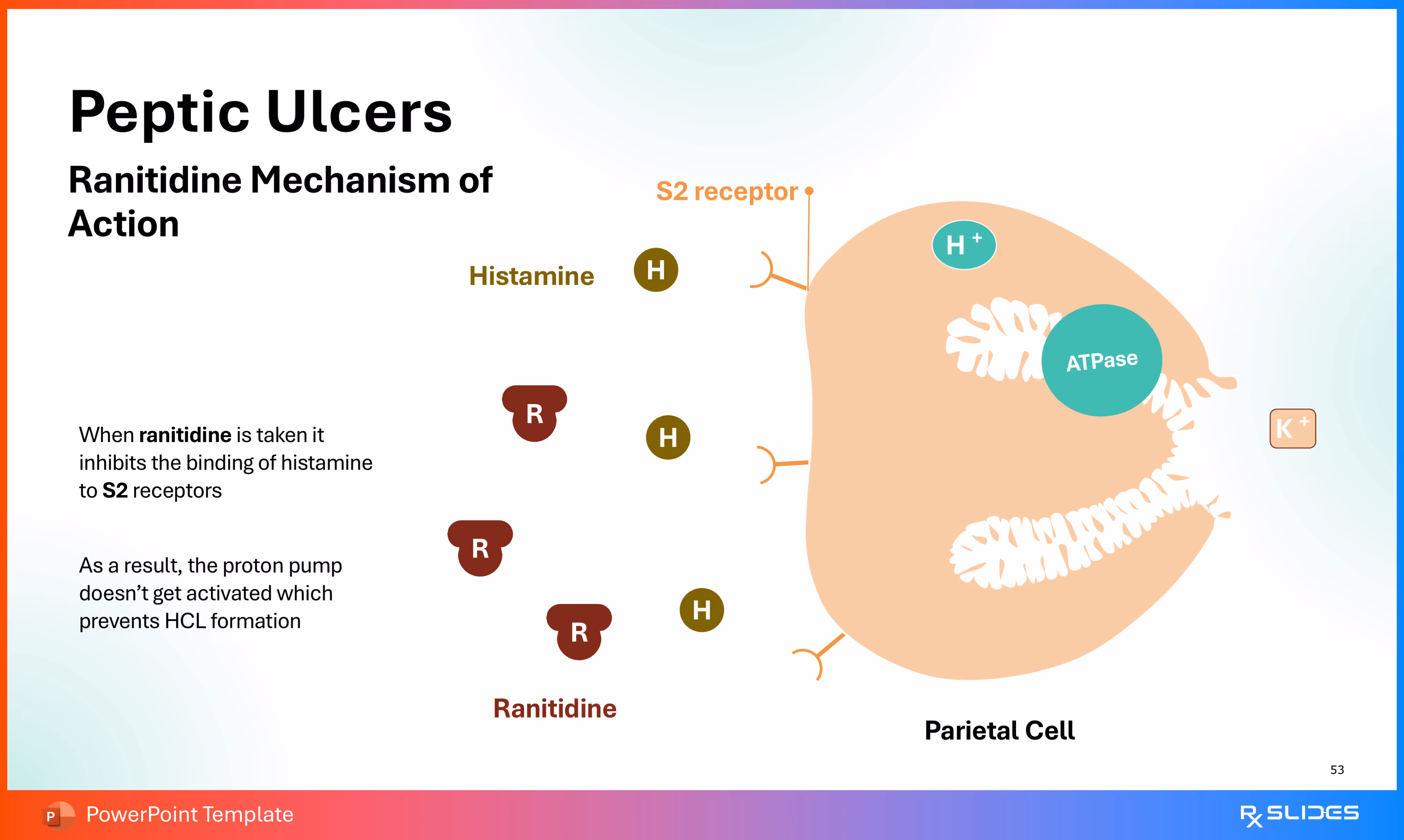
This slide concludes the molecular explanation by showing how H2-receptor antagonists, like Ranitidine, prevent acid secretion, acting as an alternative treatment to PPIs (Proton Pump Inhibitors).
- A diagram of the Parietal Cell showing the H2 receptor.
- Mechanism Details:
- When Ranitidine (represented by 'R' icons) is taken, it inhibits the binding of Histamine (H) to the H2 receptors.
- As a result of Histamine blockage, the proton pump (ATPase) doesn't get activated, which prevents the final formation of HCL.
- The illustration shows H+ being ready for secretion via the ATPase pump, but the Ranitidine molecules block the Histamine signaling pathway.
Slide 54 - Antacids for Peptic Ulcers
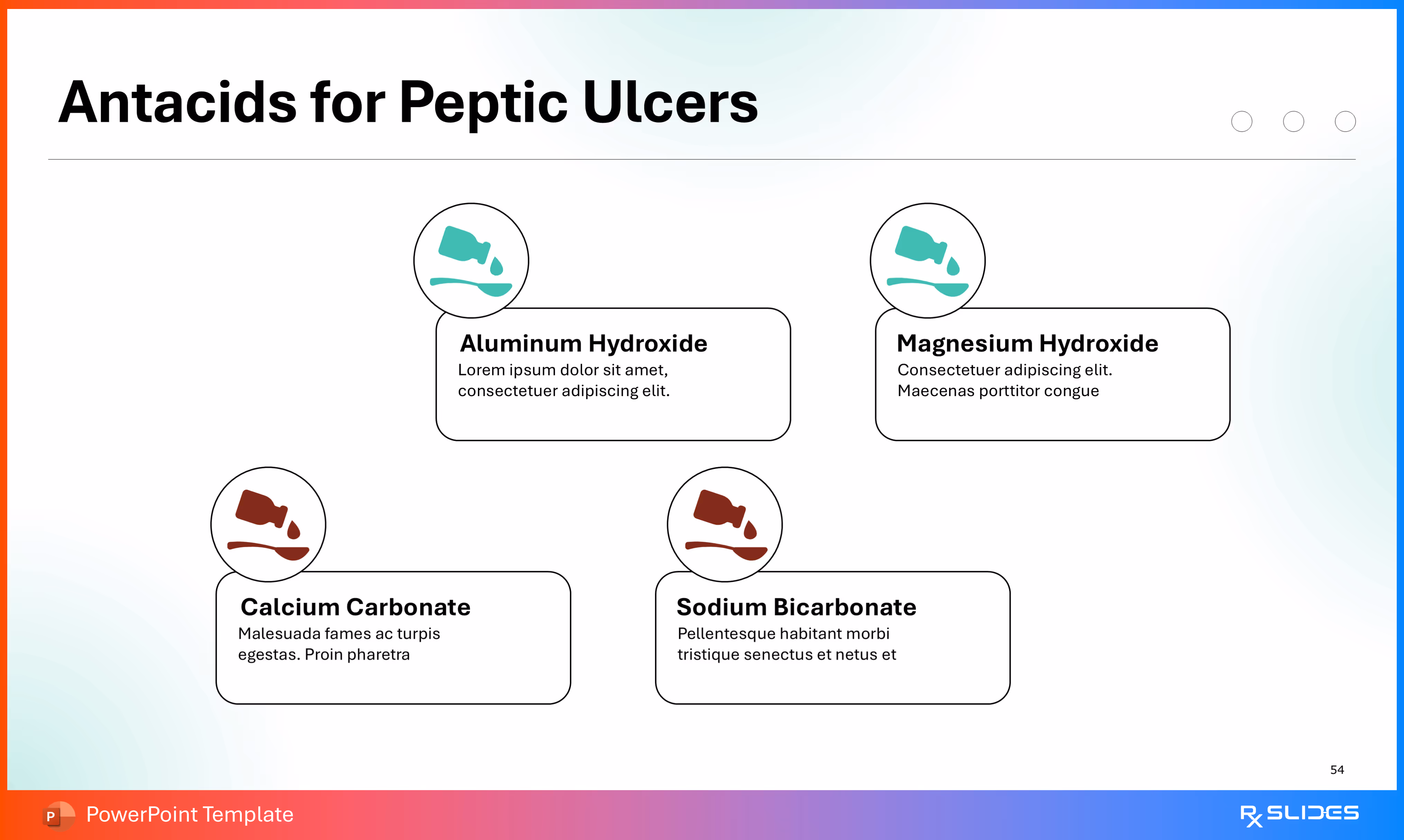
This slide provides examples of common Antacids, the fourth class of pharmacological treatment for peptic ulcers, completing the treatment section overview.
- Four common antacid ingredients are presented in a four-quadrant layout, each with an icon of liquid being poured:
- Aluminum Hydroxide
- Magnesium Hydroxide
- Calcium Carbonate
- Sodium Bicarbonate
Slide 55 - Antacids Mechanism Of Action
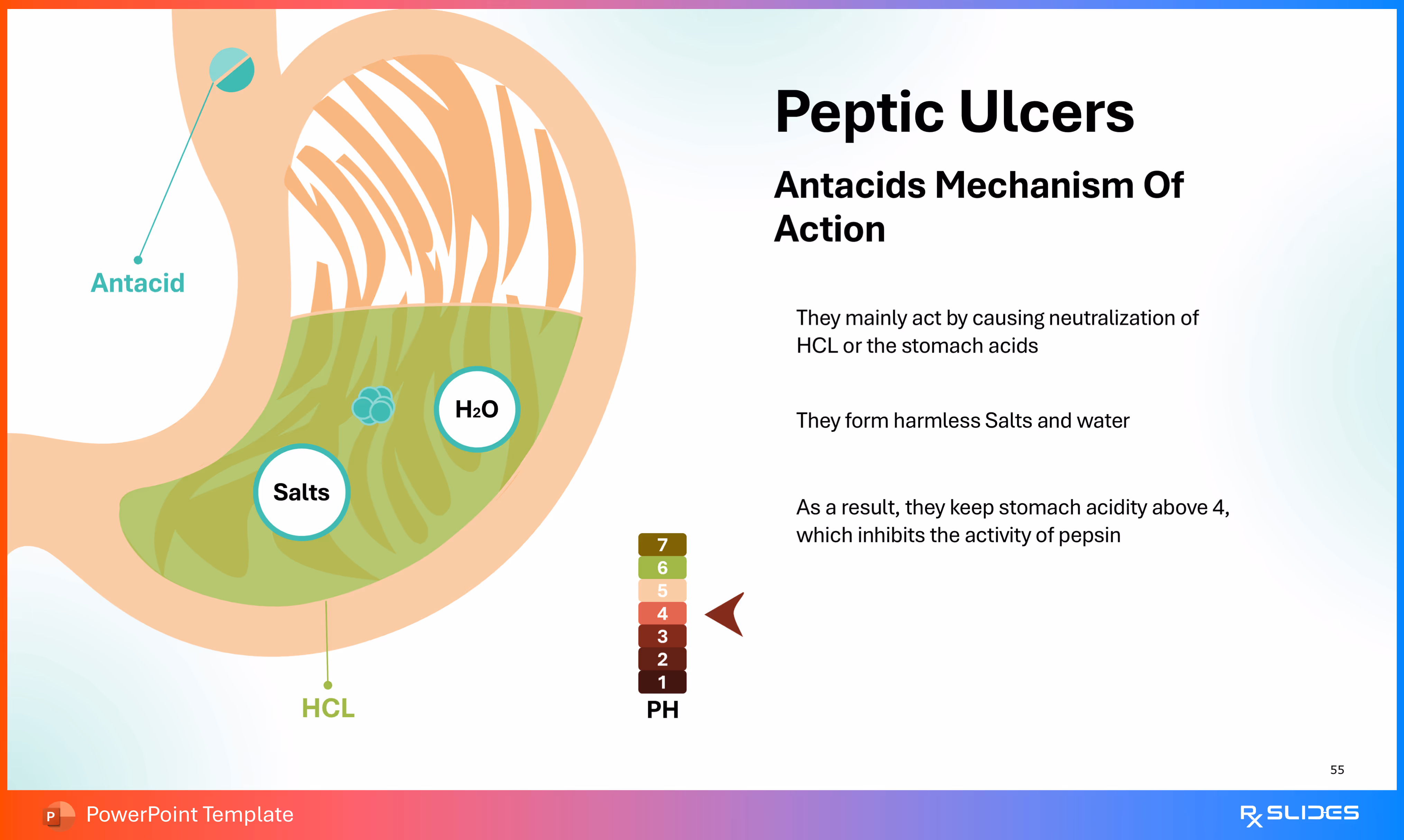
This slide explains how Antacids work to treat peptic ulcers and manage symptoms, completing the sequence of treatment mechanisms.
- Mechanism Details:
- Antacids mainly work by causing neutralization of HCL or the stomach acids.
- They form harmless Salts and water.
- As a result, they keep stomach acidity above 4 (pH>4), which inhibits the activity of pepsin.
- Visual Elements:
- A diagram of the stomach shows an Antacid entering the stomach and neutralizing HCL to form H2O and Salts.
- A vertical pH scale on the right graphically illustrates raising the pH from the highly acidic range (1-3) up toward 7.
Slide 56 - Peptic Ulcers Surgical Treatment

This is a section divider slide that introduces the topic of surgical interventions for peptic ulcers, signaling the transition from pharmacological treatment to surgical options.
- The slide features a large icon depicting surgical tools (scissors and a scalpel), emphasizing the surgical nature of the upcoming content.
Slide 57 - Peptic Ulcers Vagotomy Surgery
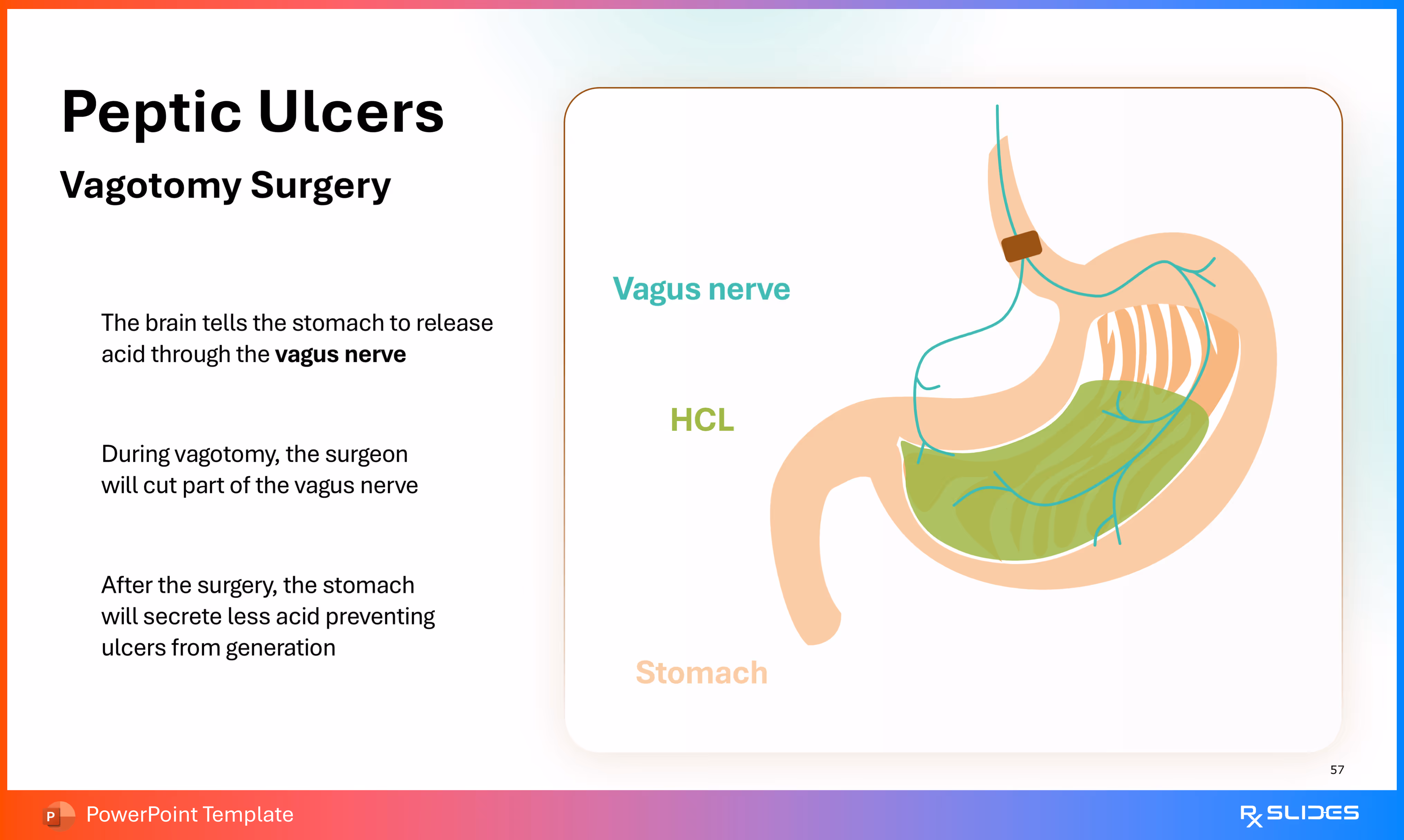
This slide explains the mechanism of a specific surgical procedure, Vagotomy, used to reduce stomach acid secretion for treating peptic ulcers.
- Mechanism Details:
- The brain tells the stomach to release acid through the vagus nerve.
- During vagotomy, the surgeon will cut part of the vagus nerve.
- After the surgery, the stomach will secrete less acid, preventing ulcers from generation.
- A diagram illustrates the stomach and the Vagus nerve running down to it, with HCL (acid) shown in the stomach lumen.
Slide 58 - Peptic Ulcers Antrectomy Surgery
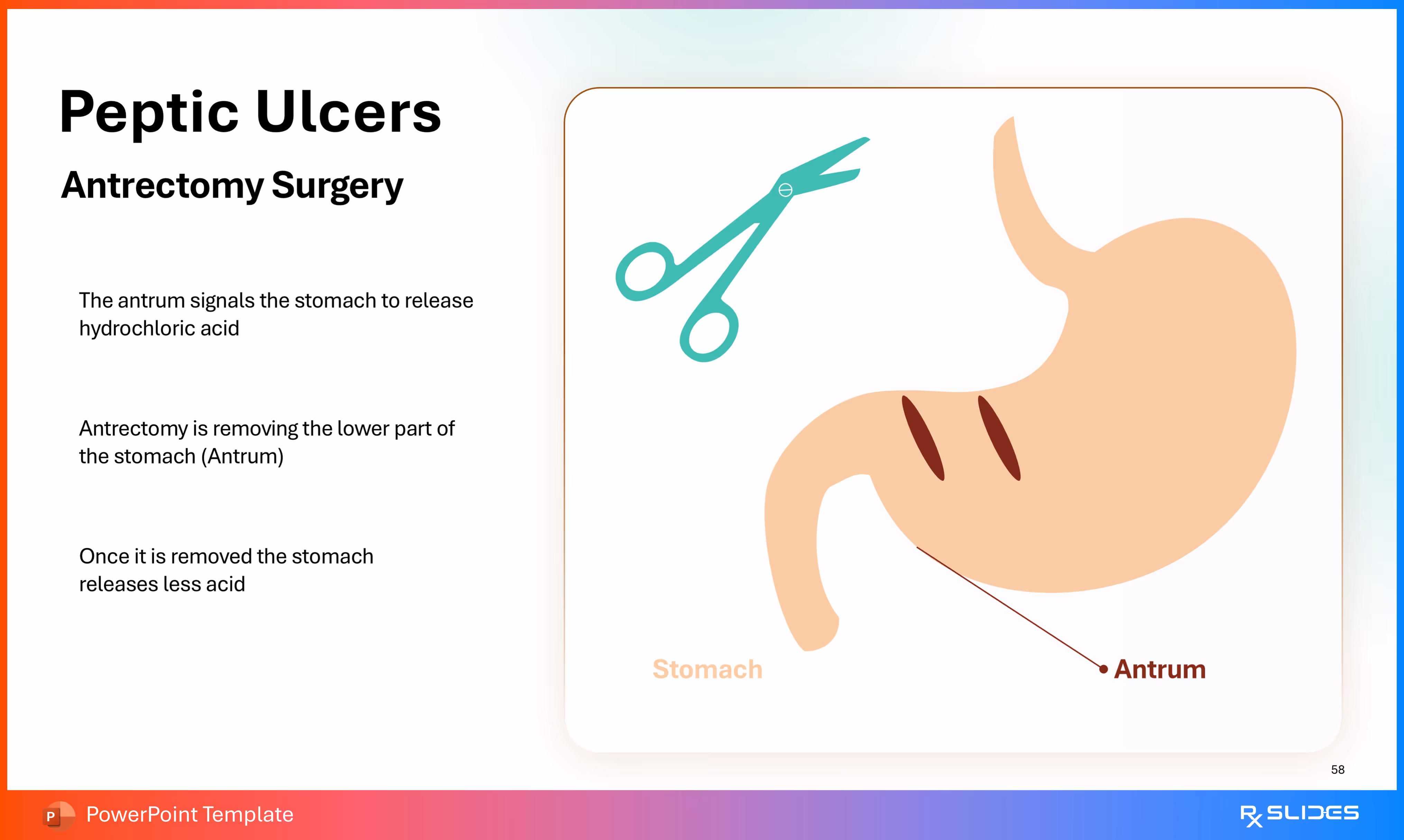
This slide explains the mechanism of the surgical procedure Antrectomy, another option used to reduce stomach acid secretion for treating peptic ulcers.
- The Antrum signals the stomach to release hydrochloric acid.
- Antrectomy involves removing the lower part of the stomach (Antrum).
- Once the Antrum is removed, the stomach releases less acid.
- A diagram of the stomach shows the Antrum labeled at the bottom, with surgical cut marks to illustrate its removal. Surgical scissors are also shown at the top.
Slide 59 - Peptic Ulcers Antrectomy Surgery (Cont. Diagram)
.avif)
- This slide provides an simplified diagram to illustrate the continuation Antrectomy procedure where The antrum signals the stomach to release hydrochloric acid
- Antrectomy is removing the lower part of the stomach (Antrum)
- Once it is removed the stomach releases less acid
Slide 60 - Peptic Ulcers Pyloroplasty Surgery
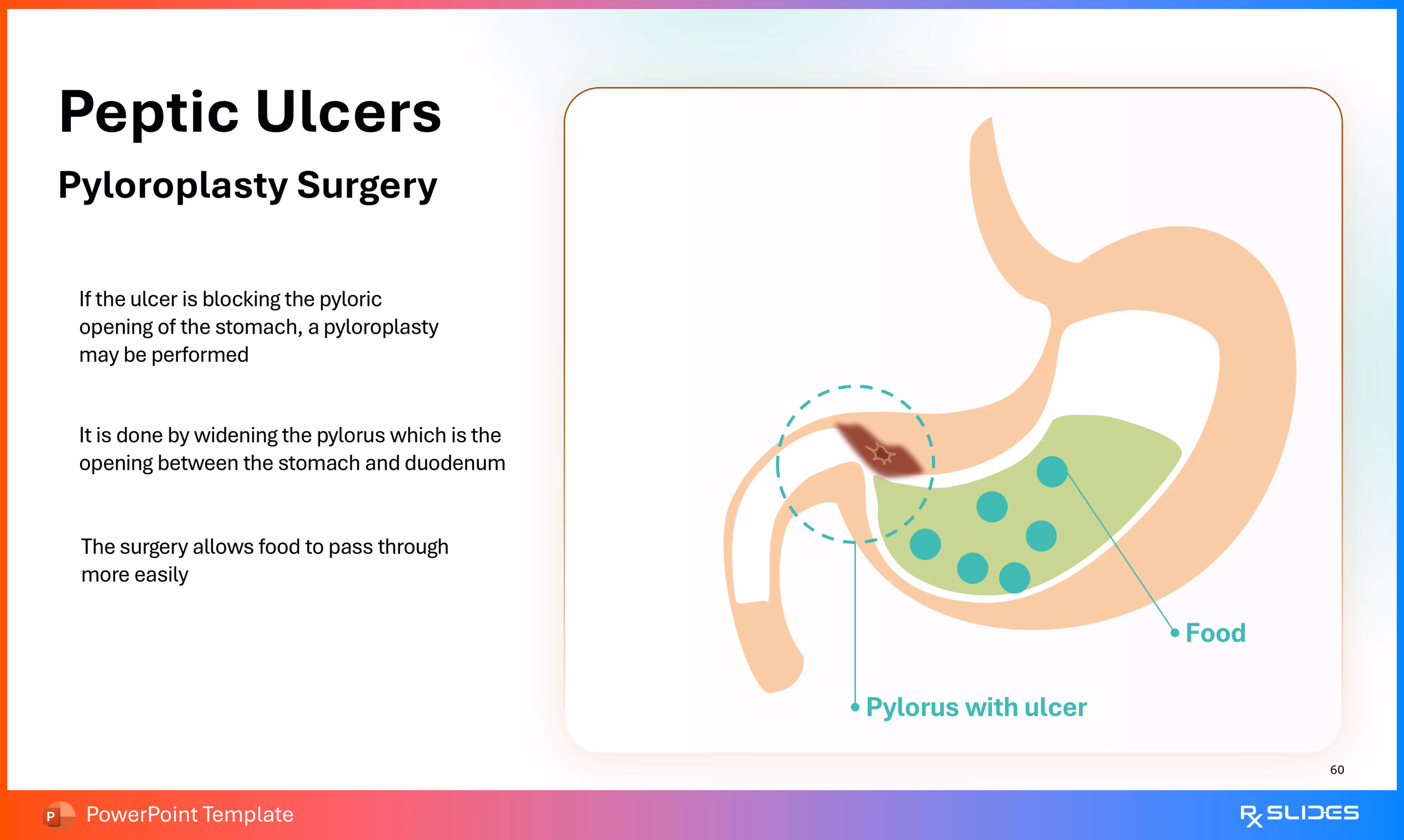
This slide explains the mechanism of the surgical procedure Pyloroplasty, which is used to address blockages associated with ulcers in the pylorus.
- Mechanism Details:
- If an ulcer is blocking the pyloric opening of the stomach, a pyloroplasty may be performed.
- It is done by widening the pylorus, which is the opening between the stomach and duodenum.
- The surgery allows food to pass through more easily.
- A diagram of the stomach shows the Pylorus with ulcer causing a blockage where Food (represented by green circles) is accumulating. A dashed circle indicates the area of widening.
Slide 61 - Peptic Ulcers Pyloroplasty Surgery cont.
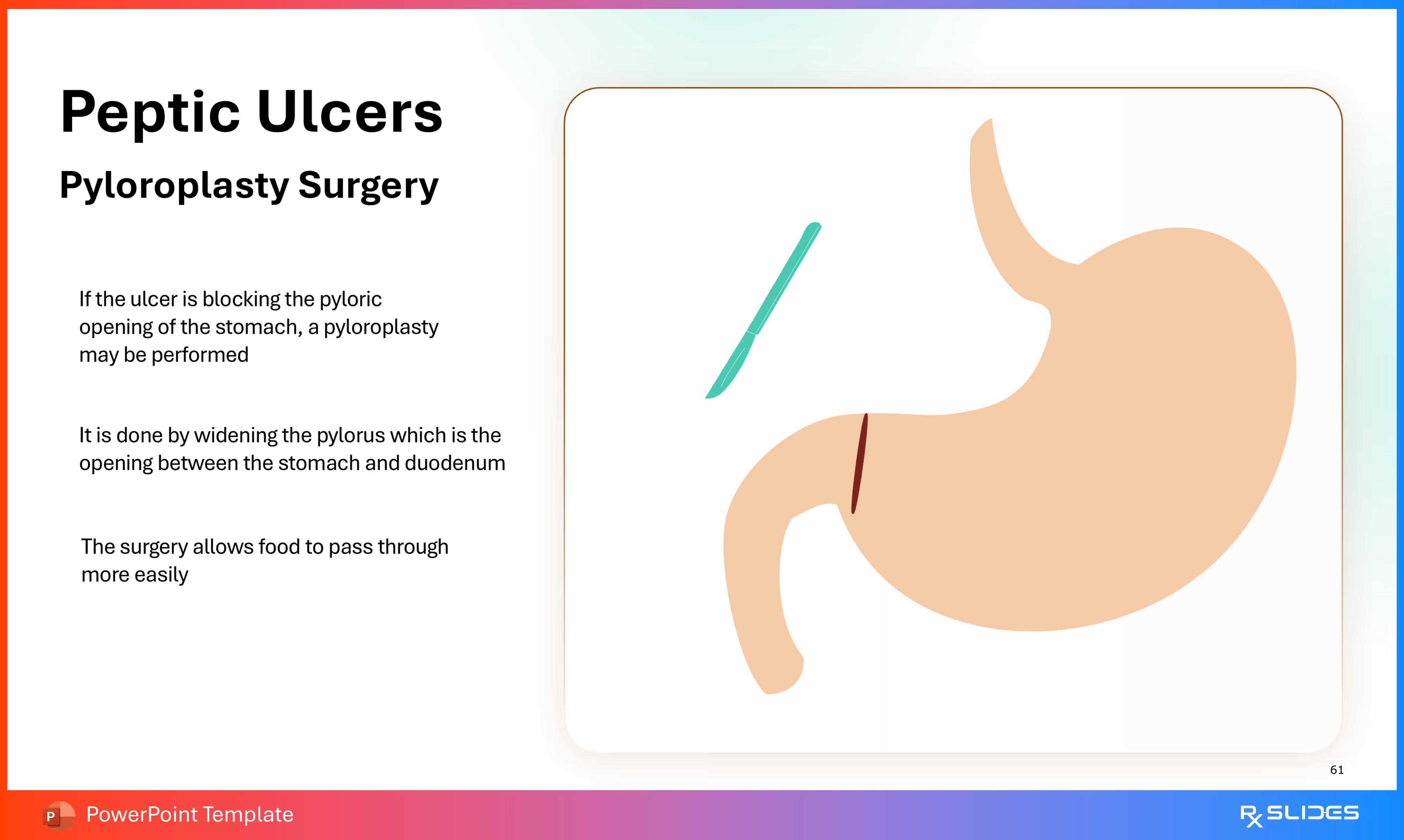
This slide provides an alternative or simplified diagram to illustrate the Pyloroplasty procedure.
- A diagram of the stomach shows an incision mark at the lower end (pylorus) and a surgical tool, illustrating the widening of the pyloric opening.
Slide 62 - Peptic Ulcers Pyloroplasty Surgery cont. 2
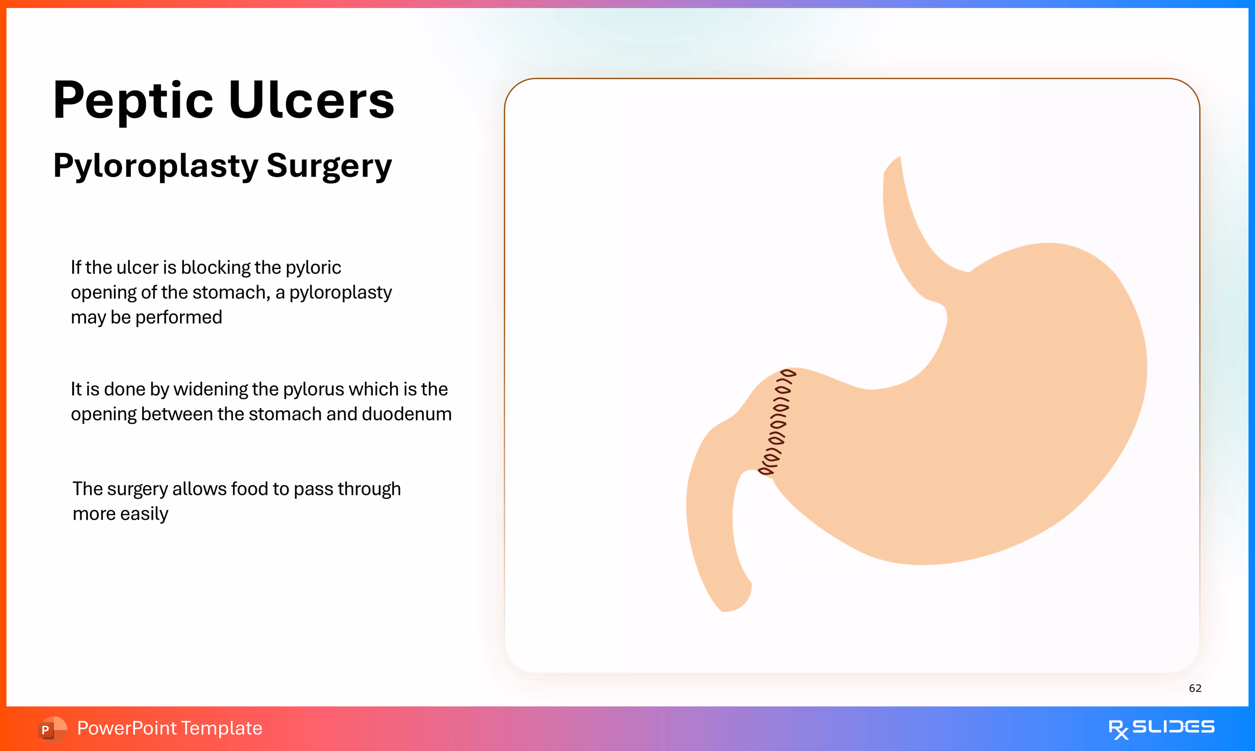
This slide provides another alternative or simplified diagram to illustrate the Pyloroplasty procedure, focusing on the surgical repair.
- A diagram of the stomach shows the anticipated repair site (pylorus) indicated by suture-like circles. This suggests the opening has been widened and closed to prevent blockage.
Slide 63 - Peptic Ulcers Complications
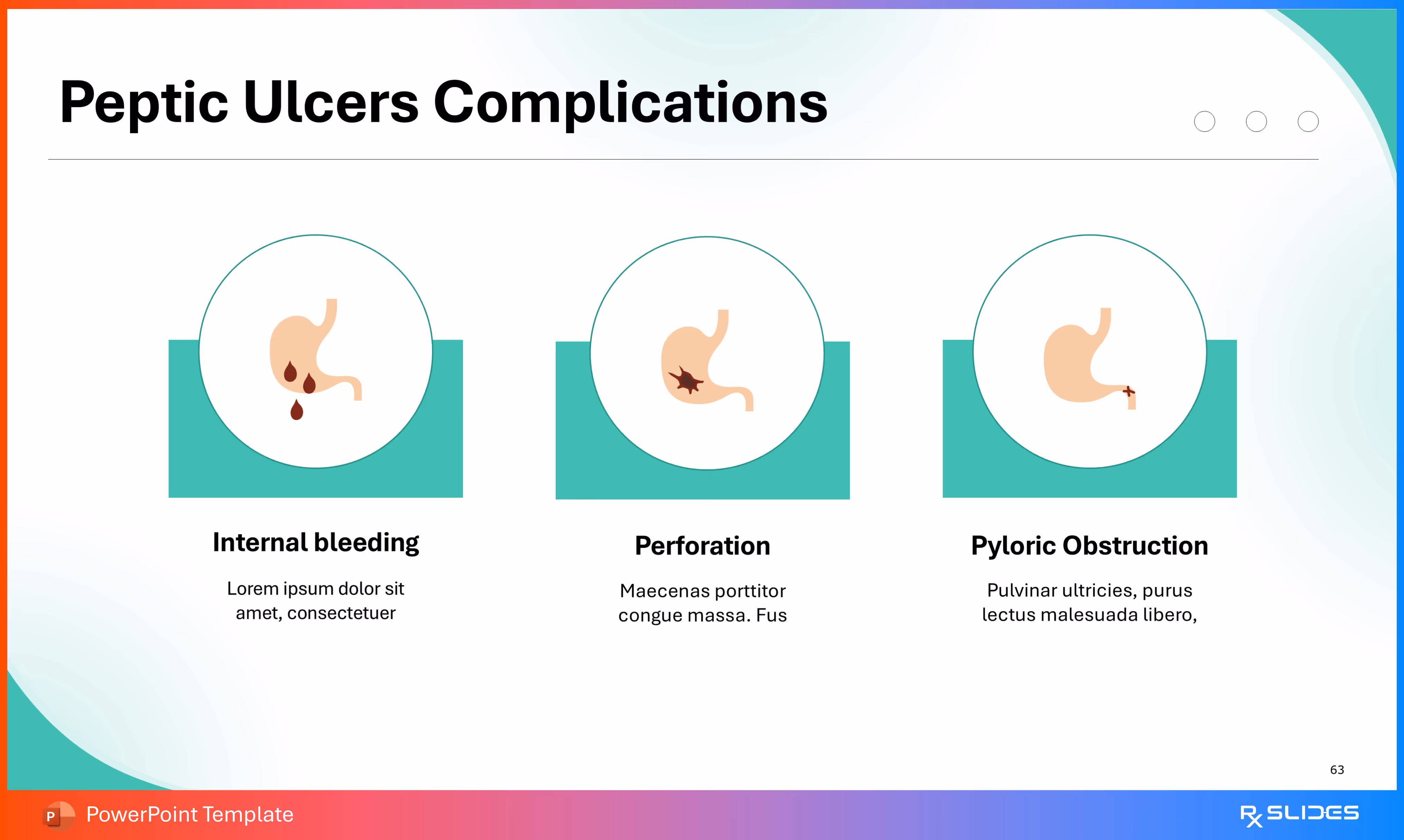
This slide outlines the three primary, severe complications that can arise from untreated or severe peptic ulcer disease.
- The slide presents three major complications in a horizontal, circular icon layout:
- Internal bleeding: Represented by a stomach icon showing blood dripping from an ulcer.
- Perforation: Represented by a stomach icon showing a star-like break in the stomach wall, indicating a hole.
- Pyloric Obstruction: Represented by a stomach icon showing the pylorus area narrowed or blocked.
Slide 64 - Thank You

This is the concluding slide for the presentation template.
Features of the Template
- 100% editable PowerPoint template.
- Editable colors, you can change according to your presentation style and company branding guidelines.