Carpal Tunnel Syndrome PowerPoint Template

.avif)
.avif)

No items found.
Carpal Tunnel Syndrome: Animated Medical PowerPoint Template
- The Carpal tunnel syndrome (CTS) PPT template is an animated medical PowerPoint template that will help you realize the full potential of your presentation.
- RxSlides include medical animations and infographics, which will attract your audience.
- You can take advantage of our demonstrated infographics to provide your audience with a dynamic presentation on carpal tunnel syndrome.
- With our Bone PowerPoint Templates, you can communicate complex information about muscles, bones, and joints to any audience.
Carpal Tunnel Syndrome PowerPoint Template Preview
Carpal Tunnel Syndrome PowerPoint Template Content
Slide 1 - Title Slide

- Clean, medical-themed opening slide with the title "Carpal Tunnel Syndrome" and space for presentation title, presenter name, institution, and references.
- Features a central illustration of the wrist anatomy, highlighting the carpal tunnel structures and setting a professional tone.
Slide 2 - Carpal Tunnel Syndrome Agenda

- Circular diagram layout illustrating the main sections of the presentation (likely Anatomy, Causes, Diagnosis, and Treatment).
- Uses numbered circles and directional arrows surrounding the central wrist illustration to clearly guide the audience through the lecture structure.
Slide 3 - Carpal Tunnel Syndrome Introduction (Section Divider)
.avif)
- Transitional slide introducing the initial section of the presentation.
- Features a stylized illustration of a hand experiencing characteristic symptoms like numbness and tingling (paresthesia) in the distribution of the median nerve, setting the context for the clinical overview.
Slide 4 - Carpal Tunnel Syndrome (Symptom Visual)
.avif)
- Visual diagram illustrating the affected area in Carpal Tunnel Syndrome (CTS).
- Features a hand with the median nerve pathway highlighted, along with visual cues (lightning bolts and wavy lines) to denote pain, numbness, and tingling typically felt in the thumb, index, middle, and half of the ring finger. Includes numbered, editable text boxes for defining the condition or listing key symptoms.
Slide 5 - Carpal Tunnel Syndrome (Ergonomic Risk)
.avif)
- Visual diagram highlighting a common ergonomic risk factor for CTS: the repetitive strain from using a computer mouse. Features an illustration of a hand clicking a mouse with visual cues (lightning bolts) denoting pain/inflammation, allowing for a discussion of occupational causes and preventative ergonomics.
Slide 6 - Carpal Tunnel Syndrome (Internal View)
.avif)
- Detailed visual diagram showing the internal anatomy of the wrist.
- Highlights the median nerve passing through the carpal tunnel beneath the transverse carpal ligament, with a visual overlay (red ring) emphasizing the area of compression and inflammation. Includes editable text boxes for defining the underlying pathophysiology.
Slide 7 - Carpal Tunnel Syndrome (Annotated Anatomy)
.avif)
- Detailed visual diagram featuring the wrist anatomy with a focus on the compressed median nerve.
- This layout includes three editable text boxes that point to the illustration, ideal for annotating key structures, defining the pathophysiology of median nerve entrapment, or listing the cardinal symptoms.
Slide 8 - Carpal Tunnel Anatomy (Detailed Cross-Section)
.avif)
- Advanced anatomical slide featuring a detailed cross-section of the wrist alongside a view of the median nerve distribution in the hand.
- Key structures are labeled, including the Flexor Tendons, Carpal Arch, Transverse Carpal Ligament, and Median Nerve. This is essential for explaining the precise mechanism of nerve compression.
Slide 9 - Carpal Tunnel Syndrome Prevalence (Section Divider)
.avif)
- Transitional slide introducing the Epidemiology and Prevalence segment of the presentation.
- Features a stylized map of the world, serving as a visual indicator for presenting global or regional data on the incidence, prevalence, and affected populations of Carpal Tunnel Syndrome.
Slide 10 - Carpal Tunnel Syndrome Prevalence (Data Layout)
.avif)
- Data-driven slide summarizing the global or regional prevalence of CTS.
- Features a world map with pin icons to visually denote research areas or high-incidence regions, alongside a numbered list for presenting specific statistical data or epidemiological findings.
Slide 11 - Carpal Tunnel Syndrome Prevalence (Bar Chart)
.avif)
- Data-driven bar chart summarizing prevalence or incidence rates across different geographical regions (represented by country map icons).
- Clearly displays and compares data points using varying bar heights and percentages, ideal for academic or epidemiological comparisons of CTS rates globally.
Slide 12 - Carpal Tunnel Syndrome Risk Factors (Section Divider)
.avif)
- Transitional slide introducing the Risk Factors segment of the presentation.
- Features a hand illustration showing symptoms (pain, tingling) to emphasize the clinical relevance, setting the context for discussing genetic, ergonomic, and medical factors contributing to CTS.
Slide 13 - Carpal Tunnel Syndrome Risk Factors (Infographic)
.avif)
- Infographic presenting four major categories of risk factors contributing to CTS: Pregnancy, Obesity, Rheumatoid arthritis, and Repetitive stress.
- Uses a circular diagram centered on an affected hand illustration, with corresponding icons for each factor to clarify systemic, pathological, and mechanical causes.
Slide 14 - Carpal Tunnel Syndrome Pathogenesis (Section Divider)
.avif)
- Section slide marking the start of the Pathogenesis (how the disease develops) section.
- Features a strong visual diagram of the hand, highlighting the median nerve pathway, to set the context for the discussion on the precise physiological mechanisms of nerve compression and damage.
Slide 15 - Anatomy of The Wrist (Labeled Cross-Section)
.avif)
- Highly detailed, labeled diagram providing an anatomical cross-section of the carpal tunnel.
- Identifies all key structures, including the Median Nerve, Flexor Tendons, Transverse Carpal Ligament, and the major carpal bones (Hamate, Capitate, Trapezoid, Trapezium, Triquetrum, Pisiform).
- This is the definitive slide for establishing the anatomical basis of Carpal Tunnel Syndrome.
Slide 16 - Hand Innervation
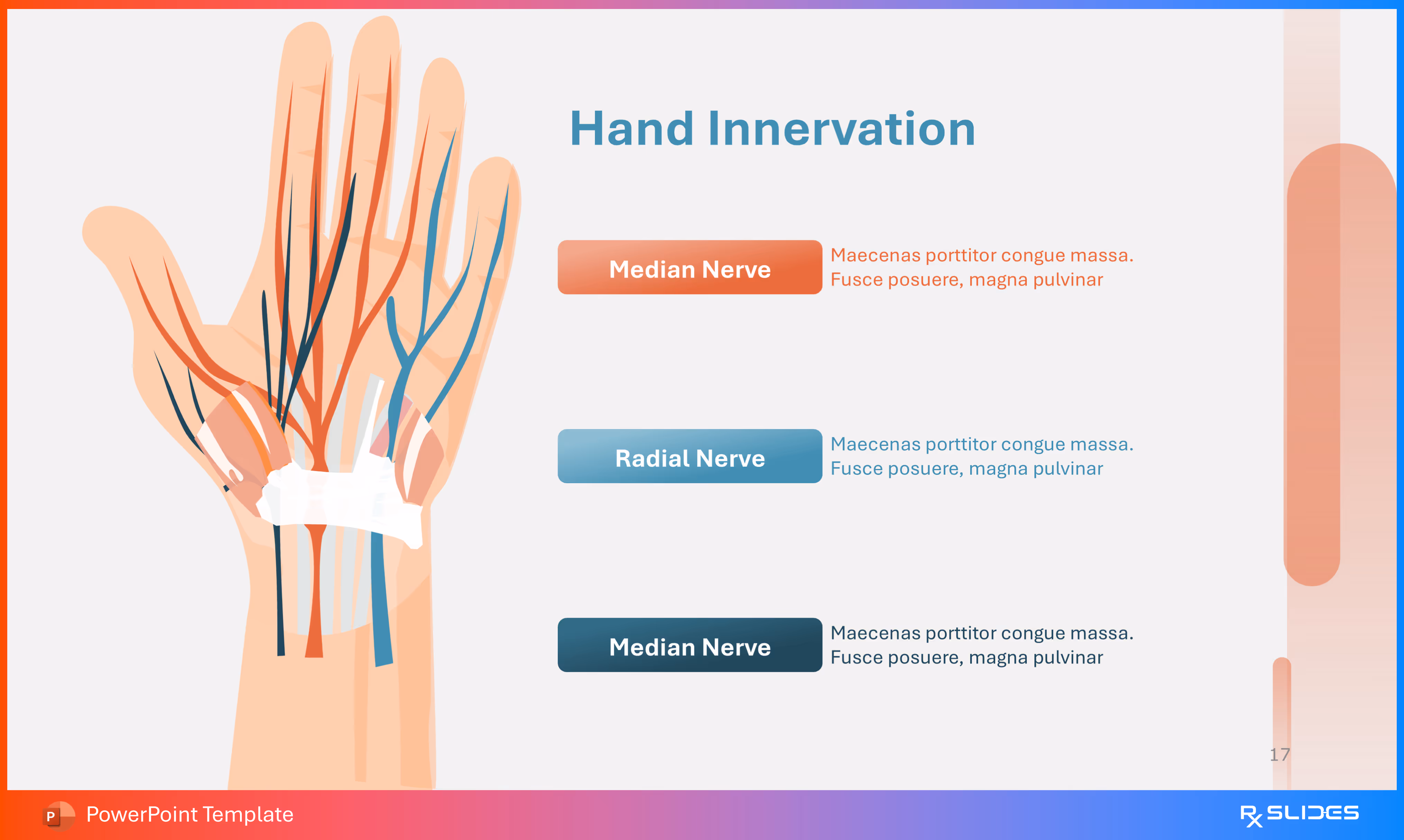
- Detailed anatomical diagram illustrating the distribution of the major nerves in the hand: Median Nerve and Radial Nerve.
- This visual is critical for contrasting the sensory and motor deficits associated with CTS (median nerve entrapment) versus other common peripheral neuropathies of the forearm and hand.
Slide 17 - Carpal Tunnel Syndrome Pathogenesis (Detailed Cross-Section with Explanation)
.avif)
- Highly detailed anatomical cross-section of the hand focusing on the compressed structures.
- The accompanying text box explains the core mechanism: inflammation of tendons and surrounding tissues creates local edema or swelling, which increases fluid and pressure in the tight carpal tunnel, directly compressing the Median Nerve and causing pain/numbness.
Slide 18 - Carpal Tunnel Syndrome Pathogenesis (Mechanism Diagram)
.avif)
- Detailed three-dimensional illustration of the hand's deep structures, visualizing the mechanism of compression.
- Highlights the Median nerve and surrounding Inflamed Tendons within the carpal tunnel.
- Includes a text box to explain that inflammation leads to swelling, which compresses the nerve, causing the characteristic numbness and pain.
Slide 19 - Carpal Tunnel Syndrome Pathogenesis (Clinical Symptoms)
.avif)
- Diagram illustrating the clinical consequences of median nerve compression.
- Highlights the Thenar Muscle (often affected by atrophy in advanced CTS) along with visual indicators for key symptoms: Paresthesia (numbness/tingling) and Dull Pain.
- Includes three editable text boxes for detailing the progression of these clinical findings.
Slide 20 - Carpal Tunnel Syndrome Symptoms (Section Divider)
.avif)
- Transitional slide introducing the Clinical Symptoms section.
- Features a stylized hand illustration highlighting the central wrist area, visually preparing the audience for a detailed discussion on the clinical presentation, subjective complaints, and physical examination findings in CTS.
Slide 21 - Carpal Tunnel Syndrome Symptoms (Circular Infographic)
.avif)
- Circular infographic detailing five major clinical symptoms experienced by patients with CTS: A shock-like sensation, Objects tending to fall off (clumsiness), Pain in hand and wrist, Numbness and tingling, and Pain being worse at night.
- This visual format is excellent for patient education and clinical lectures.
Slide 22 - Carpal Tunnel Syndrome Symptoms (Icon Timeline)
.avif)
- Horizontal, six-point timeline detailing the key clinical features of CTS: A shock-like sensation, Things tend to fall off, Pain in hand and wrist, Numbness and tingling, Pain is worse at night.
- This layout uses clear icons and flow to present symptoms in an organized and digestible manner for patients and students.
Slide 23 - Carpal Tunnel Syndrome Symptoms (Icon Grid)
.avif)
- Grid layout summarizing five major clinical symptoms experienced by patients with CTS: A shock-like sensation, Things tend to fall off, Pain in hand and wrist, Numbness and tingling, and Pain is worse at night.
- This visual layout uses distinct icons and color-coded circles for a clear, scannable overview of clinical presentation.
Slide 24 - Carpal Tunnel Syndrome Diagnosis (Section Divider)
.avif)
- Transitional slide introducing the Diagnosis section.
- Features a stylized hand illustration highlighting the wrist area, preparing the audience for a discussion on clinical tests (Phalen's, Tinel's sign) and instrumental diagnostics (Nerve Conduction Studies).
Slide 25 - Carpal Tunnel Syndrome Diagnosis (Phalen's Maneuver)
.avif)
- Detailed diagram illustrating the Phalen's Maneuver clinical test.
- Compare the Normal state with the Affected state (where wrist flexion causes increased compression of the median nerve).
- The accompanying text box explains the maneuver's procedure (flexing the wrists for one minute) and the positive result (numbness in the median nerve distribution).
Slide 26 - Carpal Tunnel Syndrome Diagnosis (Tinel's Sign)
.avif)
- Detailed diagram illustrating the Tinel's Sign clinical test.
- Shows a clinician tapping the median nerve at the wrist with a reflex hammer.
- The accompanying text box explains the procedure (tapping the transverse carpal ligament) and the positive result (reproducing tingling or pins-and-needles sensations in the median nerve distribution).
Slide 27 - Carpal Tunnel Syndrome Diagnosis (Durkan's Test)
.avif)
- Diagram illustrating Durkan's Test (Median Nerve Compression Test).
- Features a manual technique where the examiner uses their thumb to compress the carpal tunnel.
- The accompanying text box explains the procedure (manual compression for 30 seconds) and the positive result (reproduction of tingling or paresthesia), which is highly indicative of CTS.
Slide 28 - Carpal Tunnel Syndrome Diagnosis (Durkan's Test - Alternate Visual)
.avif)
- Alternate diagram illustrating Durkan's Test (Median Nerve Compression Test).
- Features a hand-drawn illustration showing the examiner's hands manually compressing the carpal tunnel over the median nerve.
- Explain the procedure: applying manual compression with the thumb for 30 seconds to reproduce the symptoms of tingling or paresthesia.
Slide 29 - Carpal Tunnel Syndrome Treatment (Section Divider)
.avif)
- Transitional slide marking the start of the Treatment and Management section.
- Features a strong visual icon of a pill and capsule, setting the context for discussing conservative, pharmacological, and surgical interventions for CTS.
Slide 30 - Carpal Tunnel Syndrome Treatment (Core Modalities)
.avif)
- Infographic summarizing four primary treatment approaches for CTS: Wear a wrist splint (conservative management), Hand exercises (physical therapy), Painkillers (pharmacological/medical), and Surgery (definitive intervention).
- This layout uses distinct icons and color-coded circles to clearly differentiate the multi-modal management strategy.
Slide 31 - Carpal Tunnel Release Surgery (Anesthetic Step)
.avif)
- Diagram illustrating the first step of the surgical procedure: Local Anesthetic administration.
- Features a hand with a syringe being injected into the wrist area.
- Explain the use of an injection to numb the wrist area, ensuring the patient does not feel pain during the operation.
Slide 32 - Carpal Tunnel Release Surgery (Incision Step)
.avif)
- Diagram illustrating the key step of making the surgical incision.
- Features a hand with a dotted line indicating the location of the small cut made during the procedure, alongside an icon of a Scalpel.
- Highlight that a small cut is made in the hand to access and release the pressure on the median nerve.
Slide 33 - Carpal Tunnel Release Surgery (The Release)
.avif)
- Detailed anatomical diagram illustrating the core goal of the surgery: releasing the median nerve from compression.
- Features an open-hand view showing the Median Nerve and the surrounding Carpal Tunnel structures, with an accompanying Scalpel icon.
- The text reinforces that a small cut is made to provide relief to the compressed nerve.
Slide 34 - Carpal Tunnel Release Surgery (Post-Procedure Summary)
.avif)
- Detailed anatomical diagram showing the surgical area.
- It illustrates the Median Nerve and Carpal Tunnel structures with text boxes summarizing the entire surgical process:
- Small cut is made in the hand.
- The carpal tunnel inside your wrist is cut so it no longer puts pressure on the nerve.
- The cut is closed by sutures and a clean bandage is applied.
- This slide is the perfect conclusion to the treatment section, walking the audience through the definitive step for nerve relief.
Slide 35 - Carpal Tunnel Syndrome Exercises (Section Divider)
.avif)
- Transitional slide introducing the Physical Therapy/Exercises segment of the treatment plan.
- Features a stylized hand icon demonstrating a specific hand or finger flexion movement (indicated by the blue arrow), setting the context for a discussion on stretching, gliding, and strengthening exercises for managing CTS symptoms.
Slide 36 - Carpal Tunnel Syndrome Exercises (Modality Breakdown)
.avif)
- Three-part grid layout detailing specific therapeutic exercise modalities for CTS:
- Finger exercise: Illustrated by hands demonstrating finger flexion/extension.
- Active range of motion: Illustrated by hands demonstrating wrist deviation or rotation.
- Tendon glides: Illustrated by a hand showing finger positioning intended to glide the flexor tendons within the carpal tunnel.
- This slide is essential for the rehabilitation and physical therapy component of the treatment plan.
Slide 37 - Carpal Tunnel Syndrome Exercises (Stretch and Strength)
.avif)
- Three-part grid layout detailing different physical therapy exercise types:
- Wrist stretch: Illustrated by hands demonstrating passive wrist extension and flexion stretching.
- Active range of motion: Illustrated by hands showing wrist circles or gentle movement.
- Wrist flexion exercise: Illustrated by a hand using a small weight (represented by a sphere) for a strengthening exercise.
- This slide concludes the comprehensive treatment and rehabilitation plan.
Slide 38 - Thank You (Closing Slide)
.avif)
- The closing slide of the presentation. Features the text "Thank You" prominently alongside a detailed, stylized illustration of the wrist and hand anatomy, highlighting the median nerve and carpal tunnel area.
- This provides a visually relevant and professional conclusion to the medical presentation.
Features of the Template
- 100% editable PowerPoint template.
- Editable colors, you can change according to your presentation style and company branding guidelines.

.avif)